
Medial Knee Joint-Space Width Linked to Osteoarthritis Outcomes
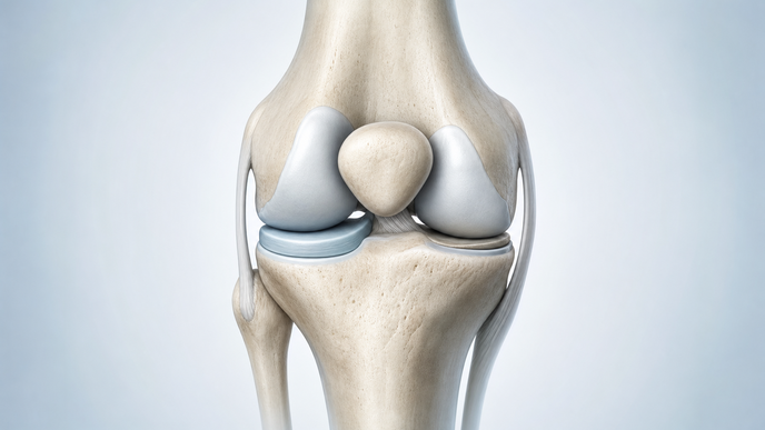
Key Takeaways
- Among UK Biobank participants with knee imaging, narrower medial minimum joint-space width was associated with knee pain, hospital-diagnosed knee osteoarthritis, and total knee replacement.
- Whole-joint minimum width showed similar associations, while lateral width showed little evidence of associations with these osteoarthritis outcomes.
- Medial, but not lateral, minimum width was associated with rheumatoid and psoriatic arthritis.
- Narrower medial and lateral widths tracked with varus and valgus alignment, respectively; varus alignment statistically mediated 16.8%, 12.7%, and 6.9% of the medial-width associations with pain, osteoarthritis, and replacement, respectively.
Investigators conducted an observational analysis of knee joint-space width in UK Biobank participants in the United Kingdom. The analysis included 53,609 participants with bilateral knee dual-energy X-ray absorptiometry (DXA) imaging. Machine-learning algorithms derived medial and lateral minimum joint-space width (mJSW); whole-joint mJSW was the smaller compartment value. Paired whole-body DXA scans provided knee alignment. Regression models accounted for both knees per participant and examined knee pain, hospital-diagnosed osteoarthritis (OA), autoimmune and crystal inflammatory arthritis, alignment, and total knee replacement. Investigators also performed sex-stratified and statistical mediation analyses.
For each standard deviation (SD) decrease in medial mJSW, the odds ratio (OR) for knee pain was 1.15 (95% confidence interval [CI] 1.12–1.18). Whole-joint mJSW showed associations with OA outcomes similar to those for medial mJSW, whereas lateral mJSW showed little evidence of such associations. Medial width was lower than lateral width on average, and most whole-joint minimum readings came from the medial compartment.
For hospital-diagnosed knee OA, the OR per SD decrease in medial mJSW was 1.29 (95% CI 1.23–1.35). Medial mJSW was also associated with rheumatoid and psoriatic arthritis, while lateral mJSW was not associated with either condition.
For total knee replacement, the hazard ratio (HR) per SD decrease in medial mJSW was 1.73 (95% CI 1.58–1.89). Narrower medial mJSW was associated with varus alignment, while narrower lateral mJSW was associated with valgus alignment. In statistical mediation analyses, varus alignment accounted for part of the medial-width associations with pain, OA, and replacement.
These observational associations and statistical mediation findings do not demonstrate that changing joint-space width or alignment changes outcomes. Measurements came from DXA scans, and the findings do not establish equivalent performance on routine knee radiographs. This UK population analysis provides neither an individual prediction threshold nor a diagnostic decision rule.
The investigators concluded that medial mJSW appears to be the most informative compartment-specific quantitative marker for large-scale population and genetically informed knee OA research. They reported that the overall compartment-association pattern did not change between inflammatory arthritis and OA clinical outcomes.
Clinician Questions
Were medial and lateral knee joint-space widths different in women and men in the UK Biobank imaging analysis?
Both medial and lateral minimum widths measured on bilateral knee DXA images were smaller in women than in men.
Can the medial knee joint-space width association be used to estimate a patient's absolute risk of knee pain?
The association was reported as an odds ratio, not an absolute patient risk. Without an absolute outcome rate or individual predictive performance, it cannot yield a patient's absolute probability of knee pain.
Recommended Reading
- For more on cartilage lesions in knee osteoarthritis: Knee Cartilage Repair: Prognostic Factors Vary by Outcome
This content is intended for healthcare professionals for educational purposes. The article and its image were developed with AI assistance and reviewed by members of the ReachMD team prior to publication. Please refer to the original source linked for the complete and most up-to-date information.