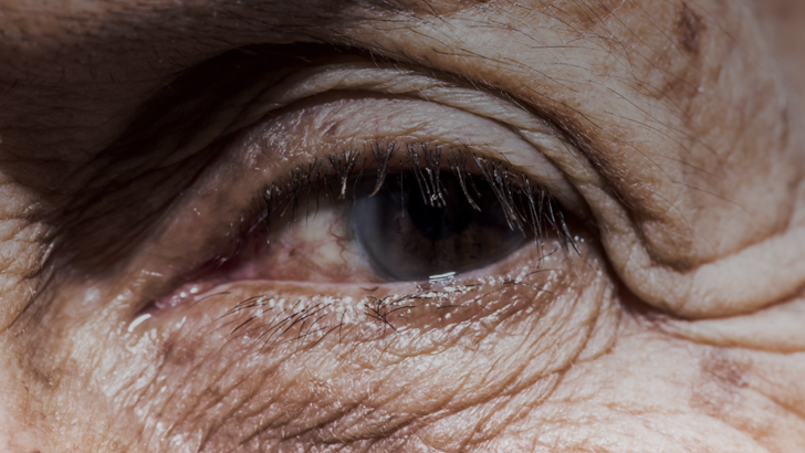
Dr. Goldberg presents two cases: an 111-year-old patient who presents for a dry age-related macular degeneration follow-up and a patient with geographic atrophy who also has advanced glaucoma and a history of retinal detachment.
GA Perspectives: Dry AMD Follow-Up and a Patient With Advanced Glaucoma and History of Retinal Detachment
Be part of the knowledge.™Register
We’re glad to see you’re enjoying ReachMD…
but how about a more personalized experience?
GA Perspectives: Dry AMD Follow-Up and a Patient With Advanced Glaucoma and History of Retinal Detachment
Restart
Resume
Transcript
GA Perspectives: Dry AMD Follow-Up and a Patient With Advanced Glaucoma and History of Retinal Detachment
closeTranscript
GA Perspectives: Dry AMD Follow-Up and a Patient With Advanced Glaucoma and History of Retinal Detachment
closeDr. Goldberg:
Thank you for joining me for this CME program provided by Evolve Medical Education. I'm Dr. Roger Goldberg. I'm in Vitreoretinal Specialist at Bay Area Retina Associates in Walnut Creek, California, and volunteer faculty at the California Pacific Medical Center Ophthalmology Residency Program in San Francisco.
Today we'll be talking about geographic atrophy and going through some cases as well to help illustrate some of these points. We know that age is the number one risk factor for AMD; after all, the name is age-related macular degeneration. And it turns out that 1/3 of adults over the age of 75 years have AMD. Individuals older than 85 years have a 10-fold higher risk of late-stage AMD, wet AMD, or geographic atrophy compared to those age 70 to 74. About 50% of the individuals with this late-stage age-related macular degeneration have this advanced atrophic form that we call geographic atrophy.
Today I'll be presenting a case of a white female woman who came to see me as a follow-up for dry age-related macular degeneration. You can see here her vision is 20/300 in the right eye and 20/80 in the left eye. And on her OCT scan, we see several important features. First of all, there are diffuse drusen present. And directly in the fovea, we see this hyper-transmission defect where we see increased penetration of the OCT laser scan penetrating deep to the retina down into the choroid and to the outer sclera. And this is a sign on OCT of RPE atrophy, of missing RPE, so you get increased penetration of the laser deep to the retina. You can also see on the infrared image, areas of atrophy in this enface view.
Here’s the fundus autofluorescence. Fundus autofluorescence is a very important imaging modality for detecting and monitoring the progression of geographic atrophy. And where there's atrophy, we see areas of hypo-autofluorescence because there's no RPE. And the RPE is what contains lipofuscin, which normally gives a natural autofluorescence to the outer retina.
Here you can see though, patchy areas of atrophy in both the right and the left eye. Interestingly, on a fundus autofluorescence, the fovea itself is also hypo-autofluorescent. So, sometimes looking at just FAF alone can make it difficult to determine whether the central fovea is involved or whether there is atrophy sparing the central fovea. For these cases, I often take a multimodal approach. Not only will I look at the fundus autofluorescence, but I also look at the OCT to look for that hyper-transmission under the fovea. And then I combine that with a visual acuity data that I'm seeing as well. And together, I use those to help guide my decision whether to treat a patient or not.
So, 20/300 in the right eye 20/80 in the left eye, should we treat this patient? And if so, which eye? Now, let me tell you a little bit more about her. Cognitively, I can tell you she's very with it. She lives alone in a single-story home, she cooks all her own meals, and she gardens every day. But by the way, she's 105 years old, pretty incredible. She's quite dynamic, a joy to take care of. And this is a patient I actually have been seeing for nearly a decade now. And these images and this vision is what I'm showing you when she's 105. But let's say she'd come in now, now that we have treatment available for patients with geographic atrophy, would I consider treating 105-year-old? I think a lot of people might hesitate to do so.
But let me tell you what's happened with her. Well, fast forward to 2023, she's now 111 years old. She's a supercentenarian. We believe she's the oldest person in the county. She still is cognitively intact, a real dynamo. She cooks, she gardens, she can no longer read, she can no longer watch TV. She, of course, doesn't drive. But now her vision has deteriorated to 5/200. So about count fingers in the right eye, and 20/350 in the left eye. And the question is, could we have helped her had we intervened all those years earlier, helped her maintain her macula and prevent or slow the growth of geographic atrophy?
Here's what she looks like now. You can see there's really diffuse RPE atrophy, diffuse geographic atrophy, the areas of hyper-transmission extend really throughout the entire macula, very large areas of atrophy on the infrared image. And when you look at the fundus autofluorescence, we see very large areas clearly now not only involving the fovea, but really taking out most of the macula. So, it can be hard to predict. And I don't think age itself should be exclusionary in the decision to determine whether we should treat a patient because it can be hard to predict how long a patient is going to live. None of us have a crystal ball, of course. And so, I think you really need to look at the individual patient factors, what their visual needs are, what their mental status is, and make a decision kind of together in conjunction with the patient.
What are some other risk factors? Well, there are some environmental and lifestyle factors as well that contribute to geographic atrophy. Smoking, of course, can be a factor in up to about a third of cases. High alcohol intake has been associated with geographic atrophy and GA progression. Certain dietary factors including the intake of large amounts of saturated fat or dietary cholesterol, and the low intake of antioxidants, vitamins, and minerals. And then finally, high sunlight exposure. For example, there's an increased rate of macular degeneration in patients who work outdoors kind of in high sunlight environments for more than eight hours a day. We also know, for example, patients who are aphakic or who had intraocular lenses implanted before the mid-80s, which were – starting in the mid-80s, intraocular lenses blocked UV light. And those patients were also at increased risk for developing age-related macular degeneration and geographic atrophy.
So, let's take a look here at another case. This is an 86-year-old, white man who is pseudophakic in both eyes, had a history of a retinal detachment in the left eye, and has advanced glaucoma – open-angle glaucoma in the left eye. Some evidence of glaucoma in the right eye, not as advanced in the right eye. A former smoker quit in 1953. Lives independently. Is 20/60 in the right eye, and 20/800 in the left eye. So, here's the right eye. And this patient has what – it can be difficult to tell on the autofluorescence here, but clearly when you look at the infrared image and the OCT, we see that the fovea, not just the foveal center but really the entire fovea, is still spared. And so, it can be difficult sometimes on an autofluorescence to tell how – whether or not the atrophy involves the fovea, again because the fovea tends to be hypo-autofluorescent on a fundus autofluorescence imaging system. So, you really have to look at the multimodal imaging, the infrared and OCT, again clearly show that the fovea is spared. Now, the fellow eye does not see well, and that fellow eye doesn't see well due to geographic atrophy in part here, but also due to the advanced glaucoma and the history of the retinal detachment. And so here, it appears on the infrared image as well as the OCT, that actually the central fovea is spared. So, you might think this patient has good visual acuity and might be a good candidate for pegcetacoplan. But again, we always have to evaluate our patients in the context of other diseases, other ocular conditions. And so, in this case, because of the advanced glaucoma and the history of the retinal detachment, this case seems to be, I think, a less ideal candidate for pegcetacoplan.
In addition to age, which is the number one risk factor for age-related macular degeneration, we know that many of our patients will have a family history of AMD. And these are patients often who are very concerned about their age-related macular degeneration because they've watched one or perhaps both of their parents lose vision over time from AMD. Sometimes they're not always sure whether they – that vision loss was from the advanced dry form, geographic atrophy, or whether it was from wet AMD in the era before we had treatment for wet age-related macular degeneration. But again, these patients are often very concerned.
And when we look at the difference between, let's say, physiologic factors, including age and certain dyslipidemias, as well as environmental factors like smoking and certain dietary changes and intakes, that it turns out that the genetics are actually responsible for about 70% of the risk of developing age-related macular degeneration. This was first actually sussed out in 2006 in a really pivotal paper in Science, which showed the role of complement factor H as a risk factor, or the genetic mutation behind some cases of age-related macular degeneration. But now many, many studies have done, including genome-wide association studies that have identified several mutations across the – across genes that impact the complement system.
In addition, we've looked at systemic levels of activated complement factors. And it turns out that patients who have age-related macular degeneration, both early stages as well as advanced stages, have elevated complement levels in their – activated complement factors in their systemic circulation. So, this really points to the role of complement being an important driver of age-related macular degeneration, and an important driver of geographic atrophy.
Of course, we've now had many studies as well, where we are inhibiting parts of the complement cascade, whether it's C3 and C3b, which is what pegcetacoplan inhibits, or C5, which is what avacincaptad inhibits. We've shown by attacking and targeting the complement system, we can reduce or slow the progression of geographic atrophy. And a lot of these complement risk factors really are genetic in origin and speak to the family history that both the patients know about and that we often ask about in clinic. Because generally speaking outside of the research setting, it's not easy to do genetic testing on these patients. There are some genetic companies out there that will do genetic testing, but these are not currently covered by insurance and so the patients have to pay out of pocket for that level of genetic detail. They are available though. But usually I just ask, do you have any family members who have AMD? And oftentimes the patients are forthcoming with that information.
Share this program on:
Choose a format
Take Post-TestSkip straight to the post-test if you have already participated in this activity
Media formats available:
0.25 credits
Completing the pre-test is required to access this content.
Completing the pre-survey is required to view this content.
Details
Presenters
Related
Comments
Overview
Disclosure of Conflicts of Interest
It is the policy of Evolve that faculty and other individuals who are in the position to control the content of this activity disclose any real or apparent financial relationships relating to the topics of this educational activity. Evolve has full policies in place that will identify and mitigate all financial relationships prior to this educational activity.
The following faculty/staff members have the following financial relationships with ineligible companies.
Roger A. Goldberg, MD, MBA, has had a financial relationship or affiliation with the following ineligible companies in the form of Consultant: AbbVie, Annexon Biosciences, Apellis Pharmaceuticals, Boehringer Ingelheim, Carl Zeiss Meditec, Coherus Biosciences, EyePoint Pharmaceuticals, Genentech/Roche, Outlook, and Regeneron. Grant/Research Support: Affamed, Annexon Biosciences, Apellis Pharmaceuticals, Boehringer Ingelheim, Carl Zeiss Meditec, EyePoint Pharmaceuticals, Genentech/Roche, Janssen, Neurotech, and Novo Nordisk. Speaker's Bureau: Apellis Pharmaceuticals, Biogen, and Genentech/Roche.
The Evolve staff and planners have no financial relationships with ineligible companies.
Nisha Mukherjee, MD, peer reviewer, has no financial relationships with ineligible companies.Learning Objectives
Upon completion of this activity, the participant should be able to:
- Review the prevalence of, and risk factors for, age-related macular degeneration (AMD) and geographic atrophy (GA)
- Compare the strengths and drawbacks of various imaging platforms and learn how GA manifests on each modality
- Identify which clinical features indicate high risk of disease progression and formulate models for adjusting practice patterns in the event of drug approval
Target Audience
This certified CME activity is designed for retina specialists and ophthalmologists.
Accreditation and Credit Designation Statements
Provided by Evolve Medical Education

Evolve is accredited by the Accreditation Council for Continuing Medical Education (ACCME) to provide continuing medical education for physicians.
Evolve designates this enduring material for a maximum of 0.25 AMA PRA Category 1 Credits™. Physicians should claim only the credit commensurate with the extent of their participation in the activity.Provider(s)/Educational Partner(s)
Evolve Medical Education LLC (Evolve) is a leader in cultivating health care for patients by educating clinical competence of the health care team. Evolve achieves this by developing and distributing high-quality, evidence-based, valid, independent CME/CE activities in a variety of learning formats. These activities are designed to increase clinician’s knowledge, skills, competence and professional performance as well as to promote professional growth, maintenance of licensure, and support quality change in care of patients.Commercial Support
This activity is supported by an independent medical education grant from Apellis Pharmaceuticals.
Disclaimer
This educational activity may contain discussion of published and/or investigational uses of agents that are not indicated by the FDA. The opinions expressed in the educational activity are those of the faculty. Please refer to the official prescribing information for each product for discussion of approved indications, contraindications, and warnings.
The views and opinions expressed in this educational activity are those of the faculty and do not necessarily represent the views of Evolve or Apellis Pharmaceuticals.
This activity is designed for educational purposes. Participants have a responsibility to utilize this information to enhance their professional development to improve patient outcomes. Conclusions drawn by the participants should be derived from careful consideration of all available scientific information. The participant should use his/her clinical judgment, knowledge, experience, and diagnostic decision-making before applying any information, whether provided here or by others, for any professional use.
System Requirements
- Supported Browsers (2 most recent versions):
- Google Chrome for Windows, Mac OS, iOS, and Android
- Apple Safari for Mac OS and iOS
- Mozilla Firefox for Windows, Mac OS, iOS, and Android
- Microsoft Edge for Windows
- Recommended Internet Speed: 5Mbps+
Publication Dates
Release Date:
Expiration Date:
Presenters

Related
 video
videoGA Perspectives: Surgeon With Blind Spots
Show more- video
GA Perspectives: An Unhappy Cataract Patient and a Patient Seeking Cataract Clearance
Show more - video
GA Perspectives: Patients Experiencing Difficulties With Reading and Driving
Show more - video
GA Perspectives: Foveal-Involving GA and Exudative AMD
Show more


Recommended
Details
Presenters
Related
Comments
Recommended
Overview
Dr. Goldberg presents two cases: an 111-year-old patient who presents for a dry age-related macular degeneration follow-up and a patient with geographic atrophy who also has advanced glaucoma and a history of retinal detachment.
Disclosure of Conflicts of Interest
It is the policy of Evolve that faculty and other individuals who are in the position to control the content of this activity disclose any real or apparent financial relationships relating to the topics of this educational activity. Evolve has full policies in place that will identify and mitigate all financial relationships prior to this educational activity.
The following faculty/staff members have the following financial relationships with ineligible companies.
Roger A. Goldberg, MD, MBA, has had a financial relationship or affiliation with the following ineligible companies in the form of Consultant: AbbVie, Annexon Biosciences, Apellis Pharmaceuticals, Boehringer Ingelheim, Carl Zeiss Meditec, Coherus Biosciences, EyePoint Pharmaceuticals, Genentech/Roche, Outlook, and Regeneron. Grant/Research Support: Affamed, Annexon Biosciences, Apellis Pharmaceuticals, Boehringer Ingelheim, Carl Zeiss Meditec, EyePoint Pharmaceuticals, Genentech/Roche, Janssen, Neurotech, and Novo Nordisk. Speaker's Bureau: Apellis Pharmaceuticals, Biogen, and Genentech/Roche.
The Evolve staff and planners have no financial relationships with ineligible companies.
Nisha Mukherjee, MD, peer reviewer, has no financial relationships with ineligible companies.Learning Objectives
Upon completion of this activity, the participant should be able to:
- Review the prevalence of, and risk factors for, age-related macular degeneration (AMD) and geographic atrophy (GA)
- Compare the strengths and drawbacks of various imaging platforms and learn how GA manifests on each modality
- Identify which clinical features indicate high risk of disease progression and formulate models for adjusting practice patterns in the event of drug approval
Target Audience
This certified CME activity is designed for retina specialists and ophthalmologists.
Accreditation and Credit Designation Statements
Provided by Evolve Medical Education
Evolve is accredited by the Accreditation Council for Continuing Medical Education (ACCME) to provide continuing medical education for physicians.
Evolve designates this enduring material for a maximum of 0.25 AMA PRA Category 1 Credits™. Physicians should claim only the credit commensurate with the extent of their participation in the activity.Provider(s)/Educational Partner(s)
Evolve Medical Education LLC (Evolve) is a leader in cultivating health care for patients by educating clinical competence of the health care team. Evolve achieves this by developing and distributing high-quality, evidence-based, valid, independent CME/CE activities in a variety of learning formats. These activities are designed to increase clinician’s knowledge, skills, competence and professional performance as well as to promote professional growth, maintenance of licensure, and support quality change in care of patients.Commercial Support
This activity is supported by an independent medical education grant from Apellis Pharmaceuticals.
Disclaimer
This educational activity may contain discussion of published and/or investigational uses of agents that are not indicated by the FDA. The opinions expressed in the educational activity are those of the faculty. Please refer to the official prescribing information for each product for discussion of approved indications, contraindications, and warnings.
The views and opinions expressed in this educational activity are those of the faculty and do not necessarily represent the views of Evolve or Apellis Pharmaceuticals.
This activity is designed for educational purposes. Participants have a responsibility to utilize this information to enhance their professional development to improve patient outcomes. Conclusions drawn by the participants should be derived from careful consideration of all available scientific information. The participant should use his/her clinical judgment, knowledge, experience, and diagnostic decision-making before applying any information, whether provided here or by others, for any professional use.
System Requirements
- Supported Browsers (2 most recent versions):
- Google Chrome for Windows, Mac OS, iOS, and Android
- Apple Safari for Mac OS and iOS
- Mozilla Firefox for Windows, Mac OS, iOS, and Android
- Microsoft Edge for Windows
- Recommended Internet Speed: 5Mbps+
Publication Dates
Release Date:
Expiration Date:
Presenters
Related
- video
GA Perspectives: Surgeon With Blind Spots
Show more - video
GA Perspectives: An Unhappy Cataract Patient and a Patient Seeking Cataract Clearance
Show more - video
GA Perspectives: Patients Experiencing Difficulties With Reading and Driving
Show more - video
GA Perspectives: Foveal-Involving GA and Exudative AMD
Show more
Facebook Comments