
Single-AAV Base Editing Restores Dystrophin in DMD Mice
Key Takeaways
- In male humanized Duchenne muscular dystrophy mice, both vectors restored more dystrophin in heart than in sampled skeletal muscle.
- Liver-detargeted MyoAAV4E-off produced markedly less observed hepatic vector DNA and no detectable target editing.
- Both treated groups had fewer centrally nucleated tibialis anterior fibers and significantly less fibrosis there, but neither had significantly less diaphragm fibrosis.
- MyoAAV4A significantly improved treadmill endurance; MyoAAV4E-off did not achieve a significant treadmill difference, although creatine kinase fell in both groups.
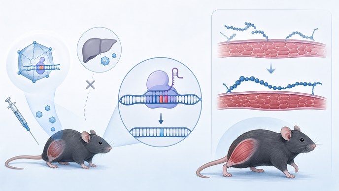
In Pien and colleagues’ preclinical study in the Journal of Clinical Investigation, investigators packaged compact SlugABE8e and an exon 45 guide ribonucleic acid (RNA) together in one adeno-associated virus (AAV) under a muscle-directed promoter. Editing the splice acceptor prompts exon 45 skipping after exon 44 deletion. Screening in human embryonic kidney 293T cells preceded AAV6 testing in exon-44-deleted, patient-derived induced pluripotent stem cell cardiomyocytes. Male humanized ΔEx44;hEx45 mice lacking dystrophin received retro-orbital MyoAAV4A or liver-detargeted MyoAAV4E-off at postnatal day 28, at 5 × 10^13 vector genomes/kg; saline-treated disease controls and wild-type mice provided comparisons. Tissue was analyzed four weeks later for genomic editing, transcript skipping, Western blot dystrophin, histology, muscle performance, serum creatine kinase, liver vector copies, and limited biochemical and inflammatory markers. MyoAAV4A and MyoAAV4E-off carried tandem and inverted cargo orientations, respectively. A high-fidelity SlugHF-ABE8e(V106W) comparison and testing of 20 computationally predicted off-target loci occurred in patient-derived cardiomyocytes, not treated mice.
Both vectors yielded measurable deoxyribonucleic acid (DNA) editing and exon 45 transcript skipping, distinct from dystrophin protein restoration. On Western blot, cardiac dystrophin reached 80.5% ± 25.2% of wild-type with MyoAAV4A versus 72.3% ± 41.6% with MyoAAV4E-off; tibialis anterior dystrophin reached 19.6% ± 6.3% versus 21.3% ± 8.8%, respectively. Restoration was lower and patchier across other sampled skeletal muscles. Patient-derived cardiomyocytes also showed editing and partial dystrophin restoration.
Liver assays found approximately 110 MyoAAV4A vector copies per diploid genome versus copies near baseline with MyoAAV4E-off; target editing was 0.5% ± 0.5% versus undetectable, respectively. Both treatments reduced centrally nucleated tibialis anterior fibers and significantly reduced fibrosis there, but neither significantly reduced diaphragm fibrosis. MyoAAV4A significantly improved treadmill endurance; the MyoAAV4E-off change was not significant. Grip-strength results differed between vectors, while serum creatine kinase fell with both. In cardiomyocytes, the high-fidelity editor had lower on-target editing at equal AAV dose; after on-target matching, it showed slightly less editing at detectable predicted off-target sites, with 1%–9% editing at a minority of sites. Serum alkaline phosphatase and bilirubin did not differ; tested tissue tumor necrosis factor (Tnf) and interferon gamma (Ifng) markers were not elevated, and gamma-glutamyl transferase was below detection.
These male-donor cells and male humanized mice do not establish human treatment outcomes; follow-up was short, and the approach targeted a specific exon 44 deletion. Different cassette orientations limit attribution of between-vector differences to capsids alone. Hepatic observations are time- and assay-specific, not proof of zero liver entry or absent systemic toxicity. Inflammatory-marker testing was not a comprehensive immunogenicity assessment, troponin was not measured, and cell-based off-target testing used selected candidates rather than genome-wide or in vivo validation. Cardiac physiological benefit and long-term durability remain unestablished.
The authors interpreted single-vector, mutation-specific editing as proof of concept: it restored dystrophin and improved selected muscle measures in humanized mice while liver detargeting lowered observed hepatic exposure. Specificity, durability, and safety remain preclinical questions.
Clinician Questions
Why does exon 45 splice-acceptor editing after a DMD exon 44 deletion produce shortened dystrophin?
In DMD with an exon 44 deletion, editing the exon 45 splice acceptor triggers exon 45 skipping and restores the gene’s reading frame. The resulting dystrophin is shorter than full-length protein; the authors cite partial redundancy among its rod domains as the rationale for retained function.
Did cardiac dystrophin restoration demonstrate improved heart function in the ΔEx44;hEx45 DMD mice?
No. The exon-44-deleted, human-exon-45-replacement DMD mice had no detectable cardiac dysfunction by fractional shortening at approximately eight weeks of age, so restored cardiac dystrophin did not establish physiological benefit. The authors note that cardiac dysfunction develops later in this model.
This content is intended for healthcare professionals for educational purposes. The article and its image were developed with AI assistance and reviewed by members of the ReachMD team prior to publication. Please refer to the original source linked for the complete and most up-to-date information.