
Pediatric EoE Shows Patchy Biopsy Findings in 15-Year Cohort
Key Takeaways
- A pediatric eosinophilic esophagitis cohort from a single center in Jerusalem showed variable symptom presentation and frequent atopic comorbidity.
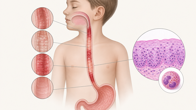
- Macroscopic abnormalities were most often distal, with furrows and exudates predominating on diagnostic endoscopy.
- Multi-level biopsy sampling identified patchy disease, including abnormal histology in some normal-appearing segments and subthreshold eosinophil counts in other sampled regions despite diagnostic disease elsewhere.
- First-line histologic remission was modest, whereas all 9 children who received dupilumab reached remission at least once during follow-up.
Investigators at Hadassah University Medical Center in Jerusalem, Israel retrospectively reviewed charts of children younger than 18 years with esophagitis diagnostic codes seen between February 2003 and October 2023 in a 15-year retrospective study. Overall, 138 children met eosinophilic esophagitis criteria, and 112 with diagnostic endoscopy and pathology available at the center formed the segment-level diagnostic subgroup. Eosinophilic esophagitis was defined as compatible symptoms plus at least 15 eosinophils per high-power field in the esophagus without an alternative diagnosis, and outcomes were followed through December 2024 with histologic remission defined as fewer than 15 eosinophils per high-power field at all sampled levels. Because this was a single-center cohort from Jerusalem rather than a U.S. cohort, the treatment patterns are local, but the sampling problem is relevant to North American endoscopic practice.
Vomiting was reported in 39% of children, dysphagia in 32%, and food impaction in 29%. Atopic comorbidity was common, lower-esophageal abnormalities were the most frequent visible pattern, and furrows and exudates were the leading endoscopic findings. Some diagnostic endoscopies looked normal overall, yet microscopic abnormalities beyond eosinophilia were still common, which reinforced the need for broad biopsy sampling.
Multi-regional sampling was performed in 100 of 112 patients, and counts below the diagnostic threshold still appeared in 21% of upper, 7% of middle, and 14% of lower samples despite diagnostic disease elsewhere. Endoscopic-histologic discordance affected 44 of 229 sampled segments and 32 of 112 patients. Histologic abnormalities were often found in normal-appearing upper and lower segments, with the upper esophagus showing the strongest discordance signal.
Histologic remission after first-line therapy was achieved in 44 of 92 patients. In the small treatment-experienced dupilumab subgroup, all 9 treated children reached remission at least once during follow-up, and 7 of 9 had done so by the first follow-up endoscopy after induction. Mild dupilumab adverse events were self-limited and included conjunctivitis and pruritus with pharyngitis.
The treatment and sampling findings came from a retrospective, single-center, uncontrolled analysis that depended on existing records and contemporary diagnostic criteria. Macroscopic endoscopic impressions were based on clinical documentation and acknowledged by the authors as subjective, and some children did not undergo repeat endoscopy, limiting confirmation of sustained histologic remission. Within those limits, the observed mismatch between visible and microscopic disease supports systematic multi-level sampling, whereas the dupilumab experience remains descriptive real-world follow-up in a small treatment-experienced subgroup.
The authors concluded that pediatric eosinophilic esophagitis in this cohort showed marked segmental variability in visible disease, relatively diffuse microscopic inflammation, and frequent endoscopic-histologic mismatch. They also reported uneven remission with conventional therapies and encouraging early experience with biologic escalation in a small group, and they called for larger prospective multicenter studies.
Clinician Questions
How was pediatric eosinophilic esophagitis defined in this Jerusalem cohort?
Pediatric eosinophilic esophagitis was defined as compatible signs or symptoms plus at least 15 eosinophils per high-power field anywhere in the esophagus without an alternative diagnosis, and histologic remission required fewer than 15 eosinophils per high-power field at all sampled levels.
Which children were included in the segment-level biopsy analysis for pediatric eosinophilic esophagitis?
The segment-level diagnostic analysis was limited to the 112 children whose diagnostic endoscopy and pathology reports were available at Hadassah University Medical Center, although 138 children met inclusion criteria overall and 26 children diagnosed elsewhere contributed follow-up data only.
What makes the dupilumab experience in pediatric eosinophilic esophagitis harder to generalize?
The dupilumab findings came from only 9 children, all treated after prior therapy failure, within a retrospective, single-center, uncontrolled cohort, so the results are best interpreted as descriptive real-world experience rather than a comparative estimate of efficacy.