
Nevus-Associated Melanoma: Dermoscopy and Invasiveness
Key Takeaways
- In a single-center Italian cohort of histologically confirmed NAM, the nevus component was dermoscopically visible in 45.6% of lesions, and invasive tumors were more common than in situ tumors.
- A dermoscopically visible nevus component was independently associated with invasive rather than in situ NAM.
- In the study’s detailed multivariable results, shiny white structures, blue-white veil (OR 8.3, 95% CI 4.5-15.4), and negative pigment network were independently associated with invasive NAM, while atypical pigment network was inversely associated.
- Larger nevus remnants were more often visible on dermoscopy, whereas deep location and ulceration were associated with lower visibility.
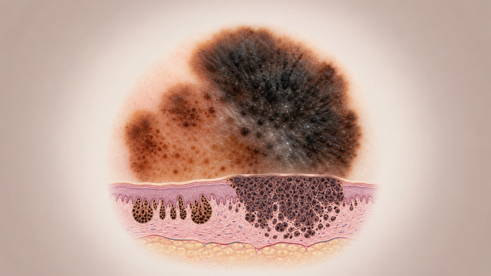
Nevus-associated melanoma (NAM) was examined in a retrospective, single-center observational study of 340 histologically diagnosed lesions treated between January 2011 and December 2024 at the Skin Cancer Centre and Pathology Unit of Arcispedale Santa Maria Nuova in Reggio Emilia, Italy, described by Spadafora and colleagues in Experimental Dermatology. Cases without high-quality dermoscopic images were excluded. Two dermatologists independently reviewed standardized polarized dermoscopic image sets while blinded to histopathologic features other than NAM histotype, and a third evaluator adjudicated disagreements over nevus visibility. An expert dermatopathologist, blinded to dermoscopic nevus visibility, reviewed nevus proportion, size, and depth on histopathology. Univariable logistic regression served as an exploratory, hypothesis-generating step before multivariable logistic regression for invasive versus in situ NAM and for visible versus non-visible nevus component, with substantial-to-excellent interobserver agreement in this middle-aged, mostly male, trunk-predominant cohort.
Among the 340 lesions, 125 were in situ and 215 were invasive. In the multivariable model, a dermoscopically visible nevus component was associated with invasive NAM (odds ratio [OR] 2.5, 95% confidence interval [CI] 1.7-3.7, p < 0.001), as were blue-white veil (OR 8.3, 95% CI 4.5-15.4, p < 0.001) and shiny white structures (OR 5.0, 95% CI 2.6-9.4, p < 0.001). Negative pigment network also remained independently associated with invasive NAM, whereas atypical pigment network and melanoma pigmentation were inversely associated with invasion.
In a separate multivariable model for dermoscopic nevus visibility, larger histopathologic nevus remnants were more likely to be recognized, with nevus size greater than 10 mm associated with higher visibility (OR 5.3, 95% CI 1.4-20.0, p = 0.013). Deep nevus location (OR 0.3, 95% CI 0.2-0.5, p < 0.001) and ulceration (OR 0.2, 95% CI 0.1-0.6, p = 0.001) were associated with lower visibility. In the adjusted model, nevus remnants measuring >2 mm showed progressively higher odds of dermoscopic visibility, whereas the 1.1–2 mm category was not significantly different from the ≤1 mm reference.
The authors noted that the retrospective, single-center Italian design and exclusion of lesions without high-quality dermoscopic images could have influenced which NAMs entered the analysis. Assessment relied on standardized clinical and dermoscopic images rather than uniform in-person examination, and dermoscopically identified nevus areas were not matched one-to-one with histopathology. Within this cohort, a nonvisible nevus component on dermoscopy did not exclude histologic nevus association. The authors suggested that small or deeply situated nevus remnants, along with melanoma-related architectural disruption such as ulceration, may help explain why some histologic NAMs appear dermoscopically nevus-negative.
Clinician Questions
How was a visible nevus component defined on dermoscopy in nevus-associated melanoma?
In nevus-associated melanoma, reviewers classified the nevus component as visible when they could identify a distinct intralesional area suggestive of residual nevus based on the overall dermoscopic appearance relative to the melanoma component and/or specific clues of a benign melanocytic lesion. Two dermatologists assessed the image sets independently, and a third evaluator resolved disagreements about nevus visibility.
How consistent were dermoscopic assessments of nevus visibility in nevus-associated melanoma?
In this Italian single-center nevus-associated melanoma cohort, agreement between independent image reviewers was substantial to excellent overall, with kappa 0.78 for nevus component visibility and values up to 1.00 for most other dermoscopic features.
Did congenital nevus association or nevus position predict dermoscopic visibility in nevus-associated melanoma?
Among nevus-associated melanomas in this series, 51 were associated with a congenital nevus, but nevus histotype did not significantly influence whether the nevus component was visible on dermoscopy. The relative horizontal distribution of the nevus component on histopathology, described as central versus peripheral, also was not significantly associated with visibility in univariable analysis.
Was blue-white veil an independent marker of occult nevus remnants in nevus-associated melanoma?
In nevus-associated melanoma, blue-white veil showed an exploratory univariable association with lower odds of a dermoscopically visible nevus component, but it was not retained in the final multivariable model. The authors therefore discussed blue-white veil as a possible masking signal rather than a confirmed independent determinant of nevus invisibility.