
Neonatal Brain MRI Abnormalities in Near-Term Infants
Key Takeaways
- In this hospitalized near-term and term neonatal ward population, abnormal brain magnetic resonance imaging (MRI) findings were reported in 35.6% of scans overall and in 34.5% of asymptomatic infants.
- Non-parenchymal hemorrhage was the most common abnormality and was reported more often with vaginal delivery, positive-pressure ventilation at birth, and hypoxic-ischemic encephalopathy.
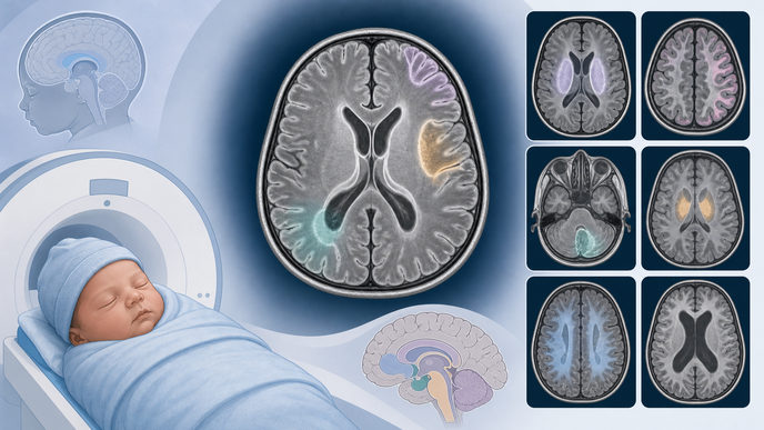
- Brain parenchymal injury was also reported, with white matter injury described as the dominant pattern.
- Subependymal cysts and congenital structural findings were less frequent, and the long-term developmental meaning of these MRI-defined injuries remained unclear.
In Hu et al. cohort of neonatal brain MRI findings, investigators retrospectively reviewed a single-center cohort from the tertiary neonatal ward at Zhangzhou Affiliated Hospital of Fujian Medical University in China, including 714 infants at 36 weeks' gestation or later who underwent MRI during hospitalization between January and December 2023. Imaging was performed on a 1.5T platform between 1 and 16 days after birth, with a median age at scanning of 5 days, using T1-weighted imaging (T1WI), T2-weighted imaging (T2WI), diffusion-weighted imaging (DWI), and susceptibility-weighted imaging (SWI). The authors classified hemorrhagic, non-hemorrhagic parenchymal, cystic, and structural findings by signal characteristics on those sequences, with hemorrhage defined by T1WI high signal, T2WI low signal, or low signal on DWI or SWI after exclusion of vascular signals.
Across the reported patterns of brain MRI abnormalities in near-term and term infants, abnormal scans were seen in 35.6% overall and in 34.5% of asymptomatic infants. Non-parenchymal hemorrhage was the leading category at 22.7%, with subarachnoid hemorrhage the most common reported subtype at 20.9%. Brain parenchymal injury was present in 14.3% of infants, and white matter injury accounted for 69.6% of parenchymal lesions.
Hemorrhage was reported more often with vaginal delivery than without hemorrhage (65.8% vs 29.2%) and was also linked to positive-pressure ventilation at birth, neurologic symptoms, and hypoxic-ischemic encephalopathy (HIE). Parenchymal injury was likewise associated with positive-pressure ventilation, neurologic symptoms, and HIE. Lower small-for-gestational-age (SGA) rates were also reported, while gestational age, birth weight, sex, maternal hypertension, gestational diabetes mellitus, hypothyroidism, and severe respiratory disease were not significantly different. Less frequent findings included subependymal cysts in 4.3%, usually bilateral or left-sided when unilateral, without measured maternal or newborn differences, and congenital variations or deformities in 1.1%, including arachnoid cysts.
The authors cautioned that this retrospective single-center neonatal ward cohort in China was enriched for perinatal risk factors and should not be read as representative of all births. Early MRI signal abnormalities may be transient, and among 591 infants with follow-up data, 33 apparent neurodevelopmental disorders were identified without a significant difference by MRI status. These findings therefore describe a Chinese tertiary inpatient population rather than a population-based estimate, and the prognostic significance of these early MRI-defined injuries remained uncertain.
In this hospitalized near-term and term cohort, the reported imaging spectrum was dominated by hemorrhage and parenchymal injury, while cystic and congenital findings were less frequent. The relationship between these early MRI-defined injuries and later neurodevelopment remained unresolved.
Clinician Questions
Which near-term and term infants underwent brain MRI in this neonatal ward cohort?
Infants in this cohort were at least 36 weeks' gestation, were hospitalized in a tertiary neonatal ward in China, and underwent brain MRI during hospitalization within the first 16 days after birth. The authors described indications including intrauterine growth restriction or small for gestational age, severe hyperbilirubinemia, perinatal asphyxia, HIE, scalp hematoma, hypoglycemia, purulent meningitis, seizures, severe respiratory distress, and fetal-ultrasound nervous system abnormalities, so the findings reflect a risk-enriched inpatient population rather than all near-term and term births.
How were hemorrhage, parenchymal injury, and cystic lesions defined on neonatal brain MRI in this cohort?
In this classification scheme, hemorrhage was defined by T1WI high signal, T2WI low signal, or low signal on DWI or SWI after exclusion of vascular signals. Non-hemorrhagic parenchymal injury was defined by T1WI high signal, T2WI low signal, or DWI high signal, while cystic lesions were defined by T2WI high signal with normal T1WI, DWI, and SWI; structural deformities were identified on T1WI and T2WI.
What brain parenchymal injury patterns were reported beyond white matter injury in these near-term and term neonates?
Beyond white matter injury, the reported spectrum included watershed-pattern injury, cerebellar hemorrhage, basal ganglia-thalamus injury, corpus callosum injury, and perinatal arterial ischemic stroke. The authors also noted that 6 infants had MRI features that could suggest bilirubin-induced brain injury.
Why did subarachnoid hemorrhage appear as the leading hemorrhage subtype in this cohort?
The authors noted that other reports have sometimes found subdural hemorrhage to predominate and suggested that the earlier MRI timing in this cohort may have contributed to the different subtype distribution. In this study, imaging was performed 1 to 16 days after birth, with a median timing of 5 days, so this remained an author interpretation tied to scan timing rather than a settled mechanism.