
Fragmentomic Liquid Biopsy Detects Early Breast Cancer and Nodal Status
Key Takeaways
- Early cancer detection showed 95% sensitivity with 78.3% specificity.
- Subtype classification across ER+/PR+HER2-, HER2+, and TNBC groups produced validation AUCs clustered around 0.89 to 0.94.
- The lymph node model showed higher negative predictive value than positive predictive value and aligned with pathology in many discordant imaging-pathology cases.
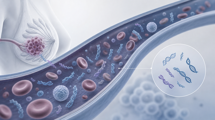
This prospective multicenter case-control study was conducted across seven Chinese institutions, including six tertiary Grade-A hospitals and one community healthcare center. The analysis set included 503 patients with breast cancer and 289 benign controls, while the Methods described 839 enrolled participants, including 550 patients with breast cancer and 289 benign controls. Among the breast cancer analysis cohort, 77.1% had early-stage disease from stage 0 through IIa, corresponding to 388 patients. Investigators used low-pass whole-genome sequencing of plasma cfDNA at about 2× coverage, with standardized processing, randomized sample processing by case/control status, and 8 to 10 mL blood collection.
For cancer detection, TuFEst produced AUCs of 0.942 in training, 0.937 in internal validation, and 0.968 in external validation, with confidence intervals reported for each cohort. The abstract reported 95% sensitivity and 78.3% specificity, and the external validation confusion matrix included 91 true positives, 4 false negatives, and 8 false positives. Those counts corresponded to a positive predictive value of 0.919 and a negative predictive value of 0.930 in the external cohort. In a distinct BI-RADS 3 subgroup with initially benign imaging assessments, the model identified 25 of 26 early-stage cancers, corresponding to 96.2% sensitivity.
A companion model, TuFEst-MS, classified ER+/PR+HER2-, HER2+, and TNBC disease, with training AUCs of 0.906, 0.925, and 0.891 and validation AUCs of 0.939, 0.925, and 0.893. Triple-positive tumors were grouped with HER2+ disease because of small numbers in that subgroup. In 21 oligometastatic patients, subtype accuracy was 85.7%, and accuracy reached 87.5% in primary-metastatic discordant cases. These findings extended the fragmentomic framework beyond detection to phenotype-level classification across primary and selected metastatic settings.
For nodal assessment, TuFEst-LN was developed in a cohort including 115 node-positive patients and showed AUCs of 0.900 in training and 0.874 in validation. Negative predictive values were 95.2% in training and 90.3% in validation, while positive predictive values were 51.5% and 54.9%, respectively. Among discordant groups, 60% of imaging-negative and pathology-positive cases were classified as node-positive, whereas 97.6% of imaging-positive and pathology-negative cases were classified as node-negative. The method was designed for radiologically detectable lesions, and limited DCIS representation constrained evaluation in precursor disease. Prospective population-based validation is still needed for radiologically occult breast cancer.