
Five CT Brain Masks Improved Cross-Center U-Net Performance
Key Takeaways
- Across 1,081 adult non-contrast head CT scans from two Chinese hospitals, investigators assessed cross-center adaptation of a public pre-trained U-Net for brain extraction.
- Fine-tuning with five local annotated scans was associated with significant Dice improvement at both centers.
- Gains beyond five scans were small and remained below the pre-specified minimal important difference, with no significant difference between the 15-sample and 75-sample models.
- Slice thickness appeared to be the dominant factor behind both baseline underperformance and recovery after fine-tuning.
- Annotation-free adaptation and from-scratch training underperformed supervised few-shot fine-tuning.
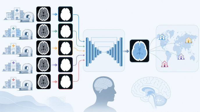
In a retrospective two-center CT brain extraction study by Sun and colleagues, adults undergoing non-contrast head CT at Liyang Hospital of Chinese Medicine and Shanghai Sixth People’s Hospital contributed one scan each. The analysis was conducted in China: 1,289 scans were screened and 1,081 were included, leaving 595 scans from Center A and 486 from Center B after exclusions for large cranial defects, severe motion, obscuring metal artifact, and duplicates. A public pre-trained U-Net, originally trained on Mayo Clinic stroke CT data, was evaluated first without local adaptation and then fine-tuned with stratified local subsets of 5, 15, 25, and 75 annotated scans from fixed 75-scan training pools, with held-out test sets of 520 scans at Center A and 411 scans at Center B. Manually delineated brain masks served as the reference standard, and the primary endpoint was the three-dimensional Dice similarity coefficient (DSC), with a minimal important difference of 0.01.
At Center A, mean DSC rose from 0.9634 ± 0.0151 to 0.9784 ± 0.0070 after five-scan fine-tuning, with ΔDSC 0.0151 [95% CI 0.0136, 0.0165], corrected p < 0.0001. At Center B, mean DSC increased from 0.9650 ± 0.0132 to 0.9813 ± 0.0078, with ΔDSC 0.0163 and corrected p < 0.0001. Later gains remained below the minimal important difference, and the 15-sample and 75-sample models were not significantly different (corrected p = 0.3764).
Five-scan performance was stable across random training subsets, pre-training remained more sample-efficient than training from scratch, and annotation-free adaptation recovered less of the performance gap than supervised local fine-tuning. At Center A, clinically unacceptable segmentations fell from 8.7% to 0.8%, and severe failures below DSC 0.90 were eliminated after five-scan adaptation. In domain-shift modeling of non-contrast head computed tomography (NCCT), slice-thickness findings in cross-center NCCT brain extraction showed slice thickness accounting for 79.6% to 90% of SHapley Additive exPlanations (SHAP) importance across baseline-performance and fine-tuning-gain models; thin-slice scans started lower and improved more. Error maps localized most discrepancies to the brain periphery and skull base, with peripheral under-segmentation improving after fine-tuning and no new systematic error regions emerging after transfer.
This retrospective study was limited to two centers, one U-Net architecture, and one public pre-trained weight set, and it assessed segmentation performance and domain-shift analyses rather than downstream diagnostic performance, treatment decisions, or patient outcomes. The authors interpreted the pattern to suggest that, for non-contrast head CT brain extraction in this setting, slice-thickness diversity may matter more than manufacturer diversity. That interpretation remains bounded to the task and acquisition context studied here.
Within this two-center segmentation study, five local annotated scans captured most of the attainable cross-center gain from fine-tuning a public pre-trained model, and slice thickness emerged as the main source of domain shift. These findings remain specific to non-contrast head CT brain extraction across the two Chinese hospital datasets studied.
Clinician Questions
Which non-contrast head CT scans benefited most from local fine-tuning in cross-center brain extraction?
Thin-slice non-contrast head CT scans, defined in the analysis as 1 mm or less, showed the lowest baseline segmentation performance and the largest recovery after few-shot fine-tuning. Slice thickness was the dominant predictor in both the regression and SHAP analyses, making it the clearest marker of which scans benefited most.
How did training from scratch or zero-annotation adaptation compare with five local annotations for CT brain extraction?
Randomly initialized U-Net models were less sample-efficient than the fine-tuned pre-trained model, and test-time batch normalization plus adversarial domain adaptation recovered less of the cross-center performance gap than supervised fine-tuning with five local annotated scans. The comparison favored retaining the public pre-training and adding a small amount of site-specific annotation rather than relying on annotation-free adaptation alone.
What counted as a clinically unacceptable or severe CT brain extraction failure in this study?
The study defined mild suboptimal segmentation as DSC below 0.95 and severe unusable output as DSC below 0.90. Using those thresholds, five-scan fine-tuning eliminated the severe failures at Center A.
What did this cross-center CT brain extraction study test, and what did it not test?
The analysis involved adults undergoing non-contrast head CT at two Chinese hospitals, one public pre-trained U-Net, manually annotated brain masks, and segmentation-performance endpoints. It did not evaluate downstream diagnostic performance, treatment decisions, or patient outcomes, and the findings were limited to this segmentation task and study setting.