
ELF3 as an Effector Gene Linking Kidney Function GWAS loci to Maladaptive Tubular Epithelial Inflammation in CKD
Key Takeaways
- At the rs10920308 kidney-function locus, kidney methylation QTL colocalization at PP4 = 0.996 and integrative mapping frameworks prioritized ELF3 as the top or sole candidate target gene.
- Across human chronic kidney disease tissue, ELF3 was enriched in injured epithelial states, especially the iPT_4 injured proximal tubule subcluster, which carried an inflammatory gene program, while stromal and immune cells showed little ELF3 signal.
- Postinjury tubular Elf3 deletion in mice was associated with fewer injury-marked epithelial cells and less leukocyte infiltration, and mechanistic assays linked ELF3 to NFκB2-centered inflammatory signaling.
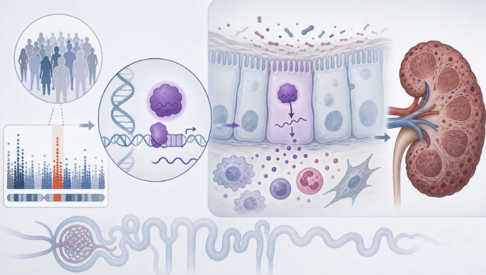
Human genetics, single-cell and spatial kidney profiling, mouse injury models, and tubular epithelial assays together localized ELF3 to injured CKD epithelium. In the human CKD atlas of 720,924 cells from 150 patients, ELF3 was concentrated in injured epithelial states, was negligible in stromal and immune compartments, and reached its highest expression in the iPT_4 injured proximal tubule subcluster enriched for IL18, NFKB1, ITGB8, CCL2, and SOX9. Spatial maps placed ELF3 expression in inflammatory epithelial regions, with minimal signal in CKD G2 and progressively higher expression in CKD G3 and G4.
Elf3 induction appeared early along the injury trajectory, with mRNA rising by 12 hours, transcript induction visible by day 4, and protein increase around day 7 in adenine injury. In the delayed-deletion experiment, mice received adenine for 1 week and then doxycycline for the next 3 weeks to ablate tubular Elf3 after injury was established. That postinjury loss was associated with lower Havcr1, Lcn2, Vcam1, and Icam1 expression, smaller KIM1-positive and Vcam1-positive areas, and less CD45-positive leukocyte infiltration, while the authors did not show reversal of established fibrosis or recovery of global renal function at the examined time point.
TNFα plus IFNγ produced the strongest ELF3 induction in primary tubular epithelial cells modeling an injured state. In that cytokine-stimulated setting rather than as a whole-kidney chromatin map, CUT&RUN identified 4536 high-confidence ELF3 binding sites and 859 conserved ELF3-bound injury-associated genes, including direct occupancy at NFκBIA, NFκB2, MAP3K14, and IL6R, with overlap across 2651 of 2731 NFκB2 target genes. Consistent with that network, Elf3 loss impaired NFκB2 nuclear localization after combined cytokine stimulation and shifted the broader inflammatory transcriptional response.
Human tissue validation placed the same program back in clinical CKD samples. In a 95-sample bulk kidney cohort, higher ELF3 correlated with lower eGFR (r = −0.253, P = 1.28 × 10⁻¹⁰) and greater fibrosis (r = 0.4, P < 2 × 10⁻¹⁶), while spatial transcriptomics localized higher ELF3 to inflammatory epithelial regions expressing NFκB2, VCAM1, and ICAM1, consistent with the ELF3 single-cell, mouse, and NFκB2 findings in CKD. The human data place the same ELF3-associated inflammatory program in progressive CKD.
Clinician Questions
Which chronic kidney disease epithelial subcluster showed the highest ELF3 expression?
In the human CKD single-cell dataset of 720,924 cells from 150 patients, ELF3 was most prominent in injured epithelial populations and peaked in the iPT_4 injured proximal tubule subcluster, which was enriched for inflammatory genes including IL18, NFKB1, ITGB8, CCL2, and SOX9; stromal and immune cells showed little ELF3 expression.
What happened after postinjury tubular Elf3 deletion in the adenine CKD model?
In the adenine CKD model, mice received adenine for 1 week before doxycycline-triggered tubular Elf3 deletion for the following 3 weeks, and Elf3 loss was associated with lower Havcr1, Lcn2, Vcam1, and Icam1 expression, smaller KIM1-positive and Vcam1-positive areas, and reduced CD45-positive leukocyte infiltration; the authors did not show reversal of established fibrosis or recovery of global renal function at the examined time point.
What evidence connects ELF3 to NFκB2 signaling in injured tubular epithelial cells?
In cytokine-treated primary tubular epithelial cells, ELF3 showed 4536 high-confidence binding sites, direct occupancy at NFκBIA, NFκB2, MAP3K14, and IL6R, and broad regulatory overlap in which 2651 of 2731 NFκB2 target genes were also bound by ELF3; Elf3 loss also impaired NFκB2 nuclear localization after TNFα plus IFNγ stimulation.
How did ELF3 expression relate to kidney function and fibrosis in human CKD tissue?
In human CKD tissue, spatial transcriptomics showed higher ELF3 expression from CKD stage G2 to G4 in inflammatory epithelial regions, and in 95 microdissected CKD kidney samples, higher ELF3 correlated with lower eGFR (r = −0.253, P = 1.28 × 10⁻¹⁰) and greater fibrosis (r = 0.4, P < 2 × 10⁻¹⁶).