
CXCR4 and PD-1 Blockade in PDAC Models
Key Takeaways
- In human PDAC tumor tissue and matched blood samples, T-cells showed a CD4-skewed pattern; in three-dimensional and patient-derived spheroid systems, infiltrating T-cells showed checkpoint upregulation within a fibroblast-rich microenvironment.
- Tumor-associated T-cells expressed more PD-1, CTLA-4, and LAG-3 than paired blood-derived cells, consistent with a dysfunctional phenotype.
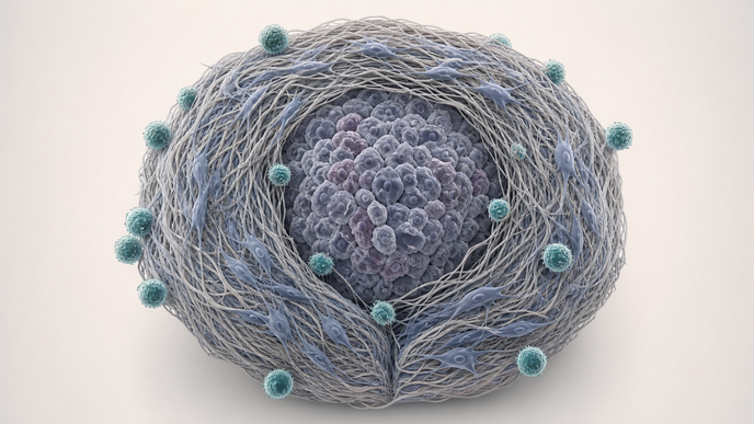
- Fibroblast-rich PSC- and CAF-containing spheroids formed an outer shell that restricted T-cell access to the tumor core and tracked with higher CXCL12 expression.
- CXCR4 blockade increased tumor-core infiltration, and PD-1 blockade partially restored effector readouts most clearly in patient-derived spheroids.
In an integrated human-tissue and three-dimensional modeling analysis, Deipenbrock et al. in Frontiers in Immunology studied immunofluorescence on formalin-fixed paraffin-embedded (FFPE) tumor sections from 10 surgically resected patients in Germany. Flow cytometry of CD4/CD8 frequencies used matched tumor and blood from five patients plus age-matched healthy-donor peripheral blood mononuclear cells (PBMC) as controls, whereas checkpoint-marker comparisons were reported in four matched patient tumor/blood samples. The models spanned PANC-1 monospheroids, PANC-1 plus pancreatic stellate cell (PSC) co-culture spheroids, and patient-derived spheroids built from primary tumor cells plus cancer-associated fibroblasts (CAF); these spheroids were infiltrated with PBMC-derived donor or autologous T-cells that had been maintained in IL-2 and activated/expanded with CD3/CD28 beads before co-culture. Immunofluorescence imaging, flow cytometry, and reverse transcription quantitative polymerase chain reaction (RT-qPCR) were used to assess spatial localization, checkpoint phenotypes, and effector readouts after AMD3100 at 4 µM for 24 hours or pembrolizumab at 1 µg/mL or 10 µg/mL for 72 hours.
Across the human tissue arm, tumor slices showed a 4.3-fold higher CD4-positive than CD8-positive T-cell frequency, and matched phenotyping found programmed cell death protein 1 (PD-1) expression was 5.7-fold higher on tumor-derived CD4-positive T-cells than on paired blood-derived CD4-positive cells. Cytotoxic T-lymphocyte-associated protein 4 (CTLA-4) and lymphocyte-activation gene 3 (LAG-3) were also higher across tumor-associated CD4-positive and CD8-positive cells, and similar checkpoint upregulation with regulatory T-cell enrichment appeared after infiltration into the three-dimensional spheroid systems. When fibroblasts were incorporated, T-cells accumulated around an outer stromal compartment instead of distributing through the tumor mass, and this peripheral pattern tracked with higher C-X-C motif chemokine ligand 12 (CXCL12) expression.
C-X-C chemokine receptor type 4 (CXCR4) blockade with AMD3100 increased tumor-core T-cell infiltration from 3% to 16.2% in PSC-containing co-culture spheroids and from 2.3% to 6.7% in patient-derived spheroids. Pembrolizumab increased interferon-gamma (IFN-γ) expression in PANC-1 monospheroids by 1.6- to 2.0-fold and by up to 5.0-fold in patient-derived spheroids, whereas PSC-containing co-culture spheroids and granzyme B/perforin readouts showed limited or nonsignificant change. Protein-level analyses also qualitatively tracked with higher IFN-γ-positive and granzyme B-positive CD8-positive cells after co-culture with patient-derived spheroids.
The evidence remains preclinical and translational, coming from human tissue analysis, in vitro three-dimensional spheroids, and ex vivo autologous models rather than clinical outcome testing. The platform was intentionally animal-free and simplified to isolate stromal and checkpoint mechanisms, which supports mechanistic dissection but does not establish clinical efficacy. Patient numbers in the tissue and matched tumor-versus-blood comparisons were modest, and pembrolizumab produced partial, model-dependent functional restoration rather than a uniform rescue across systems.
The investigators concluded that PDAC immune evasion in this system reflected two linked barriers: fibroblast-rich stroma limited T-cell access to the tumor core, and checkpoint-associated exhaustion constrained function once T-cells were present. They further described the patient-derived autologous spheroid platform as reproducing patient-specific suppression patterns and treatment responsiveness and as a tool for studying PDAC immune-evasion mechanisms and therapeutic response.
Clinician Questions
Which PDAC model preserved autologous tumor-stroma-immune interactions?
The patient-derived spheroid system combined primary pancreatic ductal adenocarcinoma tumor cells and cancer-associated fibroblasts from an individual patient with T-cells from the same patient. The investigators used it as the most patient-specific model in the study because it retained tumor heterogeneity, stromal signaling, and autologous immune crosstalk beyond the cell-line spheroids.
How was tumor-core T-cell infiltration defined in the PDAC spheroid experiments?
Tumor-core infiltration was defined by segmenting the alpha-smooth muscle actin (α-SMA)-positive shell and its interior region in three dimensions, then classifying a T-cell as a core cell when more than 50% of its segmentation intersected the interior compartment. Inside-versus-outside fractions were then quantified to measure how much of the infiltrating T-cell population reached the core.
Did pembrolizumab change PD-1 expression on infiltrating T-cells in PDAC spheroids?
After 72 hours of pembrolizumab exposure, flow cytometry did not show altered PD-1 surface expression on either CD4-positive or CD8-positive T-cells in the spheroid systems. The investigators interpreted shifts in interferon-gamma and cytotoxic effector readouts as evidence of functional PD-1 blockade rather than receptor downregulation.
What does the PDAC spheroid platform model, and what outcomes does it not test?
The PDAC spheroid platform modeled two linked barriers in pancreatic ductal adenocarcinoma: fibroblast- and CXCL12-associated exclusion of T-cells from the tumor core and checkpoint-associated functional exhaustion of infiltrating T-cells. It did not evaluate patient response, progression-free survival, overall survival, or other clinical efficacy outcomes because the evidence came from human tissue analysis, in vitro three-dimensional spheroids, and ex vivo patient-derived models.