
Carotid IPN on Ultrasound Linked to Cerebral Microbleeds
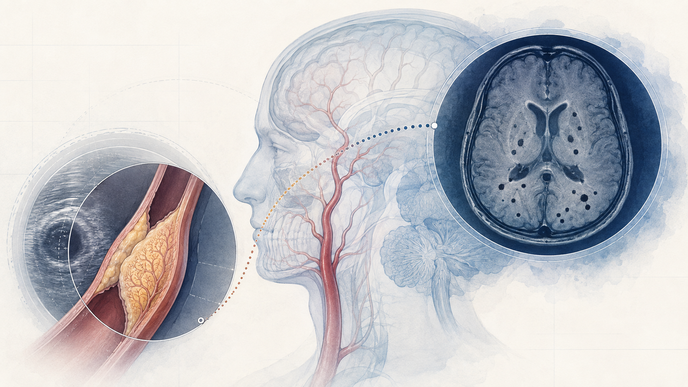
Key Takeaways
- Among neurology inpatients with carotid plaque at a single center in Hefei, China, cerebral microbleeds were detected on susceptibility-weighted brain magnetic resonance imaging.
- Higher carotid intraplaque neovascularization grade was significantly associated with greater cerebral microbleed burden.
- High-grade intraplaque neovascularization remained independently associated with overall cerebral microbleed presence after multivariable adjustment.
- In location-specific subgroup analyses restricted to isolated deep/infratentorial CMBs (n=40) versus no CMBs, high-grade IPN was associated with CMB presence, whereas no significant association was seen for isolated lobar CMBs (n=58).
Wang et al. conducted a retrospective cross-sectional single-center analysis at the Department of Neurology, Hefei Hospital, enrolling consecutive patients with carotid plaque between September 2020 and September 2024. The cohort included 493 patients, with 318 in the non-cerebral microbleeds group and 175 in the cerebral microbleeds group.
Two blinded sonographers graded intraplaque neovascularization (IPN) on a 3-point scale, with grades 0-1 grouped as low IPN and grade 2 as high IPN.
Two blinded neurologists assessed cerebral microbleeds (CMBs) on brain magnetic resonance imaging (MRI) using susceptibility-weighted imaging (SWI) and the Microbleed Anatomical Rating Scale (MARS), and categorized burden from none to at least 5 lesions, with intermediate groups of single and 2-4 lesions.
Patients with severe calcification limiting IPN assessment, prior carotid stenting, atrial fibrillation or anticoagulant use, intracerebral hemorrhage or large cerebral infarction, or major cardiopulmonary, hepatic, renal, or malignant disease were excluded. Ultrasound-defined plaque neovascularization was compared with MRI-defined microbleed burden and location.
Overall, 175 of 493 patients (35.5%) had CMBs. IPN grade correlated positively with CMB severity (rs = 0.351, p < 0.001), and high-grade IPN was independently associated with overall CMB presence (OR = 2.793; 95% CI: 1.841-4.239; p < 0.001).
Patients with CMBs also tended to be older and more often had hypertension, prior ischemic stroke, and antiplatelet exposure, while some laboratory values differed. In subgroup analysis, high-grade IPN was associated with deep/infratentorial CMBs but not with lobar CMBs, for which the model estimate was OR = 1.638; 95% CI: 0.873-3.073; p = 0.124.
The cross-sectional design does not support causal inference, and the inpatient single-center neurology setting with frequent ischemic stroke history and antiplatelet use may limit generalizability because of selection bias. Inflammatory markers such as interleukin-1 beta and interleukin-6 were not included in the regression adjustment. The authors frame carotid IPN on Angio PLUS as a noninvasive imaging marker related to cerebral microvascular injury and risk stratification rather than as proof of mechanism or treatment effect.
Higher carotid IPN grades paralleled greater CMB burden in this cohort. The association was seen for overall and deep/infratentorial CMBs but not for lobar CMBs, a pattern the authors viewed as consistent with differing microbleed phenotypes by location.
Clinician Questions
How was cerebral microbleed burden defined on MRI in patients with carotid plaque?
Cerebral microbleeds were identified on brain MRI with susceptibility-weighted imaging as small round or ovoid hypointense lesions, classified by the Microbleed Anatomical Rating Scale as lobar, deep, or infratentorial, and grouped by burden as none, single, 2-4, or at least 5 lesions.
Which cerebral microbleed locations were associated with high-grade carotid IPN?
High-grade carotid intraplaque neovascularization was associated with the combined deep/infratentorial CMB subgroup, whereas the isolated lobar subgroup did not show a significant association in the location-specific model.
Which patients do these carotid IPN and cerebral microbleed findings apply to?
These findings describe consecutive neurology inpatients at a single center in Hefei, China, who had carotid plaque on ultrasonography and underwent both Angio PLUS assessment of intraplaque neovascularization and brain MRI with susceptibility-weighted imaging. The results describe that hospital-based cross-sectional population, where ischemic stroke history and antiplatelet exposure were common, rather than establishing broad generalizability or causation.