
CA2 Microglia Track α-Synuclein in Lewy Body Disease
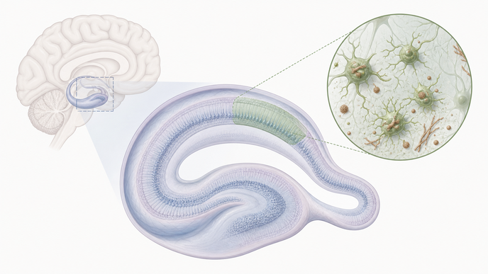
Key Takeaways
- In autopsy-confirmed Lewy body disease compared with PART controls, activated microglial marker burden was broader across hippocampal subfields.
- Within the Lewy body disease cohort, CA2 carried the heaviest neuronal α-synuclein burden and the strongest activated microglial signal.
- CA2 was the only hippocampal subfield where neuronal α-synuclein was significantly associated with both HLA-DR and CD68.
- Wider hippocampal α-synuclein distribution was associated with worse cognition and higher CA2 HLA-DR and CD68, and the CA2 signal also paralleled pathology in retrograde-connected CA4 and dentate gyrus.
In the Luna et al. Lewy body disease CA2 microglial pathology study, investigators conducted a human postmortem observational neuropathology study comparing 62 autopsy-confirmed Lewy body disease cases with 12 primary age-related tauopathy (PART) controls selected to limit confounding Alzheimer-type copathology. Validated digital histology quantified percent area occupied by neuronal α-synuclein, ionized calcium-binding adapter molecule 1 (Iba1), human leukocyte antigen-DR (HLA-DR), and CD68 across six hippocampal subfields: CA1, CA2, CA3, CA4, dentate gyrus, and subiculum. Linear mixed-effects models adjusted for demographics and age-related copathology, and cognition was related to postmortem findings using Dementia Rating Scale-2 (DRS) and Montreal Cognitive Assessment (MoCA) scores closest to death. Tissue was also classified as focal when neuronal α-synuclein was largely confined to CA2-3 and as widespread when involvement extended beyond those subfields.
Lewy body disease showed higher HLA-DR across all hippocampal subfields and higher CD68 across all subfields except dentate gyrus than PART, while Iba1 differed only slightly in CA2 and subiculum. Within the Lewy body disease cohort, CA2 carried the greatest neuronal α-synuclein burden and the highest HLA-DR signal, with CD68 also highest in CA2 and similarly high in CA3. CA2 was the only subfield where neuronal α-synuclein correlated significantly with both HLA-DR, ρ = 0.26, p < 0.05, and CD68, ρ = 0.33, p < 0.01. Neuronal α-synuclein did not correlate with Iba1 in any subfield, and the authors treated additional CD68 correlations outside CA2 as secondary.
In the subset with available antemortem cognitive data (DRS: 19 focal and 9 widespread; MoCA: 17 focal and 8 widespread), broader hippocampal neuronal α-synuclein distribution was associated with lower DRS scores. In the widespread subtype, DRS total was 97.11 ± 20.50 versus 119.16 ± 16.78 in the focal subtype, p = 0.006, and DRS memory was 15.11 ± 4.96 versus 19.63 ± 3.73, p = 0.012. MoCA trended lower without a significant difference, and CA2 HLA-DR and CD68 were higher in widespread disease. At the circuit level, CA2 microglial pathology in Lewy body disease included a correlation between CA2 CD68 and neuronal α-synuclein in CA4, ρ = 0.43, p < 0.001; HLA-DR and CD68 in CA2 also tracked α-synuclein in CA4 and dentate gyrus more broadly, but not in CA1 or subiculum. Manual CA2 neuronal counts did not differ across PART, focal, and widespread groupings, and multiplex imaging qualitatively supported greater CD68-with-HLA-DR colocalization in widespread disease.
The authors framed these findings as cross-sectional autopsy associations, not evidence that activated microglia drive α-synuclein spread or cognitive decline. Restricting the cohort to relatively pure Lewy body disease and PART sharpened the internal comparison but narrowed generalizability to mixed-pathology clinical populations; age and age-related copathology were incorporated into modeling. They also reported that the CA2 HLA-DR association was sensitive to outlier removal, whereas the CA2 CD68 association persisted modestly after outlier exclusion. Multiplex immunofluorescence came from a very small subset and was presented as supportive tissue characterization, and the retrograde-spread interpretation remained an author attribution based on correlation patterns.
Overall, the study placed activated microglial phenotypes in hippocampal CA2 alongside heavier neuronal α-synuclein burden, broader hippocampal involvement, and worse cognitive performance in Lewy body disease. The authors proposed that CA2 microglial phenotyping could refine histopathologic progression models beyond α-synuclein localization alone.
Clinician Questions
Which Lewy body disease patients do the CA2 microglial findings best apply to?
These findings come from an autopsy-confirmed Lewy body disease cohort selected to minimize clinically relevant medium- or high-level Alzheimer disease neuropathologic change, compared with cognitively healthy donors with primary age-related tauopathy and minimal other age-related pathology. That design supports a relatively pure pathology comparison and may not extend cleanly to mixed-pathology clinical populations.
How were focal and widespread hippocampal α-synuclein patterns defined in Lewy body disease?
Investigators grouped Lewy body disease tissue by visual inspection of neuronal α-synuclein staining: focal disease was relatively confined to CA2-3 with no or minimal involvement elsewhere, whereas widespread disease had substantial neuronal α-synuclein in additional hippocampal subfields beyond CA2-3. No samples showed pathology predominantly outside CA2-3.
Why did the authors interpret the CA2 pattern as compatible with retrograde intrahippocampal spread?
The authors based that interpretation on the finding that CA2 HLA-DR and CD68 tracked neuronal α-synuclein in retrograde-connected CA4 and dentate gyrus, but not in anterograde-connected CA1 or subiculum. They presented that pattern as descriptively compatible with retrograde intrahippocampal spread in Lewy body disease, not as causal proof, because the data were cross-sectional autopsy correlations.