
Blood-Cell Chromosome-Y Loss Linked to Incident AMD in Men
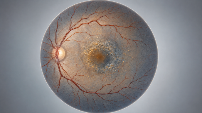
Key Takeaways
- Among UK Biobank men, blood-cell LOY was associated with greater incident AMD risk.
- MMP12 showed a significant protein signal years before the ophthalmic endpoint.
- CD16-positive circulating monocytes and resident choroidal macrophages had the highest observed LOY frequencies.
- A protein-based model discriminated incident risk in the analyzed data, without establishing screening utility.
In a longitudinal observational analysis of 223,051 men from the United Kingdom (UK) Biobank, investigators evaluated chromosome-Y loss and age-related ophthalmic disease, including incident AMD. They assessed hematopoietic LOY using median log R ratio and restricted cubic splines. Olink plasma proteomics examined a window before diagnosis, rather than a specified duration of individual follow-up. Investigators also integrated peripheral-blood and choroidal single-cell ribonucleic acid (RNA) sequencing, analyzed statistical mediation by peripheral monocyte counts, and used LightGBM to model five-year incident risk.
Investigators reported an association between hematopoietic LOY and increased incident AMD risk. The reported hazard ratio (HR) was 0.611, with a 95% confidence interval (CI) of 0.397–0.942, but the numerical reference contrast was unspecified. The estimate therefore cannot be read as the HR for LOY versus no LOY. Restricted cubic splines identified an inflection in the reported risk pattern, not a diagnostic cutoff.
Prediagnosis proteomics prioritized proteins with nonlinear trajectories, including matrix metalloproteinase-12 (MMP12), which diverged significantly 12 years before the endpoint. In integrated single-cell analyses, CD16-positive circulating monocytes and resident choroidal macrophages had the highest LOY frequencies. Y-deficient myeloid cells showed an inflammatory transcriptional phenotype with cell-type-specific IFI30 and CKB upregulation. Investigators reported significant statistical mediation by peripheral monocyte counts. The LightGBM model using prioritized proteins achieved an area under the curve (AUC) of 0.734 for incident-risk prediction.
The observational LOY–AMD association, prediagnosis protein trajectories, and statistical mediation do not demonstrate that LOY causes AMD or that monocytes transmit a causal effect. Model discrimination alone does not establish a benefit from screening. The findings remain anchored to men in the UK Biobank; applicability to other populations has not been established.
Investigators proposed a connection among somatic Y-chromosome mosaicism, immune-cell changes, and retinal disease. This remains a proposed interpretation of the observed patterns rather than an established causal pathway.
Clinician Questions
Can the chromosome-Y loss and AMD association in UK Biobank men be generalized to U.S. men?
The association between hematopoietic chromosome-Y loss and incident age-related macular degeneration was observed among men participating in the UK Biobank; its applicability to men in the United States is not established by that population.
Was the LightGBM protein model for five-year incident risk externally validated?
Investigators used prioritized plasma proteins in a LightGBM model for five-year incident-risk prediction, but external validation in an independent population is not established. Discrimination in the analyzed population alone does not demonstrate performance in another population.
Recommended Reading
- For more on dry AMD management: Illuminating Patient Selection for PBM in AMD: A Case-Based Discussion
This content is intended for healthcare professionals for educational purposes. The article and its image were developed with AI assistance and reviewed by members of the ReachMD team prior to publication. Please refer to the original source linked for the complete and most up-to-date information.