
Atopic Dermatitis Study Links Brain Glia and Immune Signals
Key Takeaways
- In adults with atopic dermatitis, LSFG-related imaging abnormalities spatially correlated with atlas-based astrocyte-, microglia-, and inflammatory gene-expression signatures relative to healthy controls.
- Left superior frontal gyrus imaging phenotypes in adults with atopic dermatitis correlated positively with IL3RA, IL13RA1, IL17RD, and IL33, while cross-modal correlations linked left superior frontal gyrus degree-centrality differences to DAT and FDOPA, and functional-connectivity differences to D2, GABAA, and NAT.
- In MC903-induced atopic dermatitis mice, prefrontal cortex neuroinflammatory changes and downregulated synaptic-plasticity programs were accompanied by lower central inflammatory readouts when Tfh13 cells were deleted and peripheral allergic inflammation was attenuated.
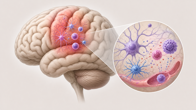
The human arm enrolled participants from 2021 through 2023 and paired resting-state fMRI degree-centrality and functional-connectivity maps with spatial transcriptomic correlation across 15,633 ranked genes. Gene-set enrichment linked the left superior frontal gyrus pattern to inflammatory response and interferon-alpha response programs, and the authors also reported KEGG signals involving asthma and staphylococcus infection, while positive correlations with IL3RA, IL13RA1, IL17RD, and IL33 framed the imaging-transcriptomic findings as a neuroimmune association rather than a causal pathway.
For the preclinical arm, investigators used a 14-day MC903/calcipotriol-induced atopic dermatitis model with prefrontal cortex RNA sequencing, RT-qPCR, histology, flow cytometry, IgE measurement, and Iba-1 immunofluorescence, using murine prefrontal cortex as the study's approximate counterpart to the human left superior frontal gyrus. The atopic dermatitis mouse prefrontal cortex showed elevated Gfap and Aif1, increased Tnf, Il6, and Cxcl10, unchanged Il1b, Ccl2, Nlrp3, and Nos2, and downregulated synaptic-plasticity-related gene sets; in Tfh13-cKO atopic dermatitis mice, the prefrontal cortex comparison versus atopic dermatitis mice showed 19 upregulated and 228 downregulated genes, alongside milder ear lesions, fewer effector CD4+ T cells and germinal-center B cells in draining lymph nodes, a downward IgE trend, downregulated interferon-alpha response, lower Aif1, Tnf, and Cxcl10 expression, and reduced anterior cingulate cortex Iba-1-positive microglial density, with Iba-1 immunofluorescence quantification reported with n=3 per group and flow cytometry and IgE analyses with n=4 per group.
The human findings were associative, while the mouse work provided mechanistic support for peripheral-central immune crosstalk.
Clinician Questions
What brain-region signal was linked to neuroinflammatory gene signatures in atopic dermatitis?
In adults with atopic dermatitis compared with healthy controls, abnormal left superior frontal gyrus activity was associated with astrocyte- and microglia-related gene-expression patterns, with enrichment of inflammatory response and interferon-alpha response pathways. The analysis linked the imaging phenotype to neuroinflammatory signatures but did not establish that the brain abnormality was caused by those pathways.
Which interleukin-related genes correlated with the left superior frontal gyrus phenotype in atopic dermatitis?
In adults with atopic dermatitis, the left superior frontal gyrus imaging phenotype correlated positively with IL3RA (r=0.586, padj<0.001), IL13RA1 (r=0.594, padj<0.001), IL17RD (r=0.651, padj=0.014), and IL33 (r=0.498, padj=0.014).
How did Tfh13-cell deletion affect central neuroinflammation in the atopic dermatitis mouse model?
In Tfh13-cKO atopic dermatitis mice, attenuation of peripheral allergic inflammation was accompanied by lower interferon-alpha-response signaling in prefrontal cortex, reduced Aif1, Tnf, and Cxcl10 expression, and lower anterior cingulate cortex Iba-1-positive microglial density, alongside milder skin findings, fewer effector CD4-positive T cells and germinal-center B cells, and a downward IgE trend. The study described these changes as concurrent with reduced peripheral allergic inflammation rather than as a treatment effect.
Recommended Reading
- For more on atopic dermatitis comorbidities: Allergic Comorbidities in Atopic Dermatitis: What the Evidence Shows
- For more on real-world atopic dermatitis: Atopic Dermatitis in the Real World
- For more on eczema diagnosis and treatment: Infant Eczema: Diagnosis, Treatment, and Parent Education