
AO-OCT Detects Cell Loss Despite Clinically Stable Glaucoma
Key Takeaways
- Treated glaucoma eyes deemed clinically stable on standard global measures nevertheless showed ganglion cell layer soma loss on AO-OCT.
- Soma density loss differed between more and less diseased hemifields.
- AO-OCT detected progression at more sampled retinal sites and earlier than local OCT thickness or visual field pattern deviation.
- Regional OCT thinning was significant in more diseased hemifields despite stable global indices.
In a longitudinal observational study in Science Advances, investigators followed one eye each from five patients with glaucoma and five age-matched healthy volunteers. Clinical stability meant no statistically detectable progression on standard global measures; glaucoma participants continued prescribed pressure-lowering drops. Four patients had six AO-OCT sessions over three years; the fifth had two sessions 4.7 years apart, and volunteers had two sessions 4.5 ± 0.3 years apart. Six macular sites per eye were sampled at 3°, 6°, and 12° temporal eccentricities above and below the horizontal midline. Investigators used vascular landmarks for longitudinal registration and manually reviewed automated ganglion cell layer (GCL) soma segmentation. They matched local clinical OCT GCL thickness and visual field pattern deviation to those sites. Progression required a statistically significant negative temporal slope; only the four patients with six visits contributed to the site-level comparison.
Annual GCL soma density declined approximately 6.86% in more diseased glaucoma hemifields and 4.16% in less diseased hemifields, versus 0.27% per year in healthy volunteers. Despite stable global indices, local clinical OCT showed significant GCL thinning in more diseased hemifields. Thinning in less diseased hemifields did not differ significantly from healthy controls, and visual field pattern deviation showed no significant longitudinal trend in either glaucoma hemifield.
At study end, AO-OCT GCL soma density detected progression at 42% of 24 retinal locations in four glaucoma patients, versus 17% by local clinical OCT GCL thickness and 8% by visual field pattern deviation. These proportions describe sampled sites, not patients.
Under the investigators’ negative-slope rule, AO-OCT detected location-level progression 0.74 years earlier than local clinical OCT GCL thickness and 1.49 years earlier than visual field pattern deviation. Remaining GCL somas also enlarged. Earlier statistical detection does not establish improved outcomes from changing treatment.
The small cohort, restricted AO-OCT field, slow acquisition and motion sensitivity, fixation-related loss of overlapping sites, and differing visit intervals across modalities constrain interpretation. Segmented GCL somas include displaced amacrine cells; individual retinal ganglion cells were not unequivocally identified or tracked over time. AO-OCT-guided treatment changes have not been shown to improve outcomes, and technical demands currently limit widespread clinical use. The authors consider cellular imaging a potential glaucoma progression biomarker pending larger prospective, multisite validation.
Clinician Questions
Why was visual field pattern deviation matched to AO-OCT macular sites in glaucoma?
Localized visual field pattern deviation reflects focal functional deficits while adjusting for diffuse loss from media opacity, making it more comparable with small AO-OCT macular regions. Investigators also accounted for anatomical displacement between ganglion-cell receptive fields and photoreceptor locations when mapping visual field data; the methods do not measure the same cells.
Why might ganglion cell layer thickness differ from cellular loss in glaucoma?
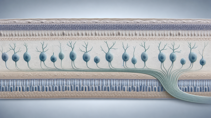
Clinical OCT measures bulk layer thickness, whereas AO-OCT segments GCL somas. Investigators observed a disproportionate decline in soma volume relative to overall layer volume, enlargement of remaining somas, and an increased noncellular fraction. The authors considered tissue remodeling a possible explanation, not an established mechanism.
This content is intended for healthcare professionals for educational purposes. The article and its image were developed with AI assistance and reviewed by members of the ReachMD team prior to publication. Please refer to the original source linked for the complete and most up-to-date information.