Transcript
Announcer:
You’re listening to ReachMD.
This episode of Living Rheum, titled “Future Imaging Modalities in Spondyloarthritis,” is sponsored by Novartis Innovative Medicines US Medical Affairs. The host and speaker have been compensated for their time. This program is intended for health care professionals.
Here’s your host, Dr Ethan Craig.
Dr Craig:
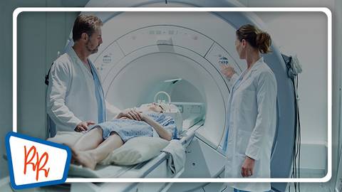
In this episode, I’m excited for the opportunity to look into the future. If you’re someone that’s been listening to this whole series and wondered if there’s anything beyond the worlds of magnetic resonance imaging and ultrasound for diagnosis of spondyloarthritis, then this episode is for you. While none of these modalities are in widespread practice, it does appear there may be some appealing options on the horizon that are coming up, including things like whole-body MRI and positron emission tomography scanning for diagnosis of SpA. In this episode of Living Rheum, we will speak with an expert on imaging in SpA and get a feel for some of the possibilities that might be out there.
This is ReachMD, and I’m Dr Ethan Craig, and there’s no one I’d rather have join me to discuss this topic than our guest, Dr Walter Maksymowych. Dr Maksymowych is a professor of medicine at the University of Alberta and he’s chief medical officer of CARE Arthritis. Dr Maksymowych, thanks for being here today.
Dr Maksymowych:
It’s a great pleasure, Ethan.
Dr Craig:
So, I think in talking about the future here, it might be a good starting point to think about the present a little bit, and maybe ask you to outline why we’d be thinking about any further imaging modalities. In some way, MRI and ultrasound seem perfectly fine. So, what do you see as the major limitation to our current options?
Dr Maksymowych:
Well, that’s a great question, Ethan, and MRI and ultrasound are indeed wonderful imaging modalities. And they’re certainly far better – MRI is far better than radiography for evaluating the sacroiliac joint because radiography is just so insensitive, and we really do not want to wait until we see changes on radiography before starting treatment of patients.
But MRI does have its problems, nevertheless. So, in children especially, interpretation of the sacroiliac joint is really quite challenging. It’s a very different scenario from adults, and that’s because of the growth plates and their irregularity of the cortical margins in children. And so, radiography is exceptionally challenging to reliably interpret in children.
And MRI can still be a problem—conventional MRI—and structural lesions like erosions in adults can be difficult. We’d really like the MRI to really look as well-defined as a CT scan, where we’re evaluating the joint margins, and it’s nice to know that, in fact, MRI has advanced to the point now, with artificial intelligence, that we can create an MRI picture that looks like a CT scan. And this was recently approved by the FDA, this technology, and we call it Bone MRI.
Dr Craig:
That’s fascinating. I wonder, you know, another technology that’s already out there, albeit somewhat limited fashion, is that of whole-body MRI. Do you see this as playing a new role in the future, and if so, what’s keeping it from wider adoption?
Dr Maksymowych:
Indeed, that’s a great question. Whole-body MRI is a technology that increasingly we’re exploring to get a better picture of what’s happening throughout the skeleton. And of course, it gives us the opportunity to simultaneously evaluate both the axial skeleton and peripheral joints and structures such as entheses, because we do want to know to what degree patients have both axial as well as peripheral inflammation.
What we don’t know at this point yet, because we’ve just started doing evaluations with whole body MRI, is how much of a problem is this? In patients with psoriatic arthritis, we expect to see more peripheral joint inflammation together with axial inflammation, and we perhaps underestimated axial inflammation in patients with psoriatic arthritis. Conversely, in patients with axial spondyloarthritis, we may have underestimated how much peripheral inflammation there is. So, it really would be nice if we had a technology that would inform us about inflammation at all of these locations. And so, psoriatic disease is certainly one subset of patients that we’d be very interested in, and it really is a technology that’s evolving. The main challenge is that this scanning protocol takes about an hour to complete, and you can well imagine this may not be comfortable for patients trying to lie still in a magnet so that we can do adequate imaging.
Dr Craig:
I think that’ll induce claustrophobia in even the most tolerant patient, yeah. You know, one area beyond MRI that’s of some interest is positron emission tomography, PET, scans, especially for its possible role in providing some in vivo imaging of metabolic pathway. You know, we’ve seen prior studies looking at metabolites like 18-flouride being taken off into actively remodeling bone, or 18F-fluorodeoxyglucose that’s taken up into synovial tissue, both of these could give us a different view on processes actually taking place in SpA. Does this seem like a technology that may actually come to prime time down the line?
Dr Maksymowych:
It’s moving in that direction, I think, Ethan. So, PET-CT, PET-MRI, are very much imaging modalities of great interest because they again may enhance our sensitivity, our ability to detect joint pathology early in the course of disease, especially before we encounter structural damage.
So, for example, PET-CT does offer the ability to study bone metabolism, and some recent, very interesting data presented at the American College of Rheumatology has also given us information about a biomarker that tells us about the presence of activated fibroblasts. And these have been shown in patients with active inflammation with psoriatic arthritis and have also been shown to demonstrate changes within the time frame, around 12 to 16 weeks, of a placebo-controlled trial in patients receiving a biologic therapy. So, this is really important that we can demonstrate change in such a short timeframe, and this, of course, informs us also about the pathogenesis of disease.
We need to understand to what degree these sorts of changes are early indicators of structural damage. What does this tell us about our ability to predict structural damage? And if we intervene, and we exert a positive change on these findings, these early findings on PET-CT and PET-MRI, is that beneficial in terms of its impact on preventing structural damage?
Dr Craig:
Finally, I’m interested in not only the imaging modalities, but whether who’s reading these images could change in coming years. There’s recently been an interest in artificial intelligence and deep learning algorithms for radiology studies. Do you see AI, do you see ChatGPT replacing us for reading these studies in the coming years, and if not, where could you see it fitting into this field?
Dr Maksymowych:
That’s a very interesting question, Ethan, and certainly there is a lot of interest and research ongoing with AI. And there are already a lot of applications in imaging, but I think we’re not quite there as far as prime time for interpretation of findings in the sacroiliac joint for diagnostic evaluation. So, what we have to bear in mind is that AI tends to work very well when it is possible to readily draw regions of interest, for example, around a bright area of inflammation that clearly differentiates from the healthy bone marrow. And then AI can be very good at discriminating between healthy and normal bone marrow, and can even quantify the extent and degree of inflammation.
The problem with the bone marrow in the sacroiliac joints and spine of adults is that the marrow signal can be heterogenous. It varies with age, and it varies with gender. And especially in spondyloarthritis, it is highly variable because we not only see the bright signal from inflammation, but we also see dark signal from new bone formation – sclerosis. So, we have this very heterogenous picture, and it’s difficult to discriminate abnormal from normal bone marrow. And so, we need to feed the AI system with lots of imaging reports and data from experts and interpretation of sacroiliac joints before we can really generate effective, reliable, and reproducible AI algorithms that are ready for prime-time diagnostic evaluation.
Where it may work is in AI being able to determine what is, in fact, a completely normal scan of the sacroiliac joints so that these are not channeled to the MSK radiologist, and all the equivocal or abnormal scans are sent to the MSK radiologist for further interpretation. So that might be the first way in which AI is beneficial.
Dr Craig:
And outside what we’ve talked about already, are there any other areas of interest you’re watching for the future of imaging in SpA?
Dr Maksymowych:
Well, I think something that could be very helpful globally for rheumatologists and radiologists to consider is low-radiation CT of the sacroiliac joint and spine. So, CT scanners, of course, are very widely available, more widely available than MRI, and the level of radiation of CT now of the sacroiliac joints is really very low indeed. Very comparable, in fact, to a chest x-ray. And so, it still gives us a high-resolution picture that allows us to interpret the joint margin to see, for example, if there’s an erosion or if there’s ankylosis. So, low-radiation CT is certainly better than conventional radiography, and even may be helpful in interpreting an MRI scan that may be a little bit doubtful. So this is an additional technology that enhances what we currently have.
Dr Craig:
That’s a great way to round out our discussion on this topic. I wanna thank my guest for joining us today, to help us think about some of the upcoming imaging modalities for diagnosis of spondyloarthritis. Dr Maksymowych, it was a pleasure speaking with you today.
Dr Maksymowych:
Thank you so much, Ethan. I really appreciate the opportunity.
Announcer:
This industry podcast was sponsored by Novartis Innovative Medicines US Medical Affairs. If you missed any part of this discussion or to find others in this series, visit reach-M-D-dot-com-slash-living-rheum.
This is ReachMD. Be part of the knowledge.