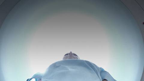
Pre-treatment MRI can eliminate unnecessary diagnostic or surgical procedures for children with suspected musculoskeletal infections. Host Dr. Jason Birnholz and Dr. J. Herman Kan, assistant professor of radiology at Vanderbilt University Medical Center, specializing in pediatric and adolescent radiology, discuss the application of MRIs and the results of his recent study which showed that a significant number of surgeries could be avoided with early MRI evaluation. Tune in to hear the valuable role MRI plays in the evaluation of musculoskeletal infection.
ReachMD
Be part of the knowledge.™Register
We’re glad to see you’re enjoying ReachMD…
but how about a more personalized experience?
Pediatric Musculoskeletal Infections: The Role of MRI
Restart
Resume
You are listening to ReachMD XM 160, the Channel for Medical Professionals. Welcome to Advances in Medical Imaging, a program discussing the latest innovations in clinical radiology and imaging technologies. Your host is Dr. Jason Birnholz, director of diagnostic ultrasound consultants in Oak Brook, Illinois.
Magnetic resonance imaging has become the standard for revealing anatomy and pathology of the musculoskeletal system. What is the role of MRI in children with suspected osteomyelitis or septic arthritis? With me today is Dr. J. Herman Kan, assistant professor in the department of radiology and radiological sciences of the Vanderbilt Children's Hospital and Vanderbilt University in Nashville. Today, we are discussing musculoskeletal MRI in children emphasis on osteomyelitis.
DR. JASON BIRNHOLZ:
Well Herman, thank you for joining us.
DR. J. HERMAN KAN:
Thank you for asking me for this interview.
DR. JASON BIRNHOLZ:
Well, I wonder if you might tell our listeners why MRI is so well suited to musculoskeletal imaging.
DR. J. HERMAN KAN:
The benefits of MRI is that there is no ionizing radiation so the child does not get exposed to radiation that you get with a CT or with plain radiography and the soft tissue and marrow delineation of MR is faster period than that you can get from any additional type of imaging such as computer tomography. MRI is very valuable for musculoskeletal imaging in children for those 2 reasons.
DR. JASON BIRNHOLZ:
When you are looking at acute neuromuscular conditions in children, are there any special requirements for equipment?
DR. J. HERMAN KAN:
The major requirement is for those children who require sedation and our principle is that if it's a boy, if they are 8 years and younger they need sedation and if they are girl, if 5 years and younger where they will need sedation. So, the main issue is the sedation factor. The other issue is making sure that when you refer a patient for musculoskeletal MR imaging is that you are referring them to a center that routinely does children. The major issue with that is that if you refer them to a place that doesn't typically deal with children, they won't have the special coils that are tailored for children, meaning specifically small surface coils that we would need or volume coils that are tailored for the small body habitus and that is important because they adequately delineate soft tissues and marrow abnormalities in children, you really need to have tailored equipment so that you can get superior imaging.
DR. JASON BIRNHOLZ:
Well, I suppose it is not always clear to our referring physicians that there are several ways to image data and several ways to analyze it and display it and that part of the job of the radiologist is to optimize the exam even before it begins to interpret the study. Are there some particular interpretive pitfalls peculiar to MRI in children?
DR. J. HERMAN KAN:
Well, the main pitfall and what you need to children is that we are imaging epiphyseal cartilage as well as physeal cartilage which are not present in adult patients and in general, especially for the preschool children the small size of the patients require us to really have a firm understanding of the normal maturation of process and the normal marrow changes that happen with development and in terms of things that are potential pitfalls for MR imaging in children, there is a normal progression of how marrow goes from red marrow to yellow marrow and if you are not familiar with how the marrow changes happen, you could potentially call marrow edema when in fact it's just normal red marrow and then similarly if you are unfamiliar with the normal ossification centers as well as how epiphyseal cartilage undergoes ossification, you may misconstrue normal developmental changes of the epiphysis for pathology when in fact it's just the normal developmental changes that happen with development.
DR. JASON BIRNHOLZ:
Let's turn into the case of osteomyelitis with septic arthritis. What's the particular clinical scenario that might prompt consideration of these conditions in a child?
DR. J. HERMAN KAN:
In general, when we are presented with a case of suspected infection, there is the Kocher criteria which is well used by many pediatricians, emergency physicians, and orthopods and the Kocher criteria are that the sed rate or CRP has to be a certain level, there has to be a white count and temperature greater than 38.5 and then nonweightbearing and based on the Kocher criteria, for instance if you have 1 of the 4 criteria, I believe there is perhaps like a 5 or 10% chance of the child having a septic arthritis, but once you go to 2 or greater of the Kocher criteria, then there is a much higher incidence of infection and so usually it's not for just 1 of the 4 Kocher criteria, it's usually when there is 2 or greater Kocher criteria where the patient is referred for us to imaging and what happens first is that they get plain radiography or ultrasound and when plain radiography is normal, I mean there is not a factor that is causing the symptoms and if we can actually image the joint, we will do it by ultrasound first and if there is a joint effusion, then the patient may end up getting aspirated, but if the ultrasound and plain films are nondiagnostic or normal, that's when the child usually is referred for MR imaging.
DR. JASON BIRNHOLZ:
What about CT by the way, does it have a role in this?
DR. J. HERMAN KAN:
CT has a very limited role when there is no history of trauma. CT plays a great role because it can be done fast and there is no need to sedate the child, especially if you have a multi-role detector CT, but in terms of looking for infection, CT does not provide the same type of soft tissue and marrow delineation that MR may afford and so it's not recommended that a child, if there is a question of septic arthritis or osteomyelitis, get a CT. They should get plain films, ultrasound if it's a joint that can be ultrasounded and then if those are negative, then they should really be referred for MR imaging.
DR. JASON BIRNHOLZ:
Well, are there ultrasound signs that would lead you to diagnosis of osteomyelitis?
DR. J. HERMAN KAN:
Not for osteomyelitis, but for indirect signs. So, I will never make the diagnosis of osteomyelitis based on ultrasound. However, especially in the hip as well as the knee, what ultrasound may afford is that it can actually delineate whether there is a joint effusion present or not and when there is a joint effusion present, then the orthopod may either aspirate the joint or if he has a high clinical concern that is not just septic arthritis, but osteomyelitis, the patient may be referred for MR imaging, so ultrasound really just looks for joint effusion and what ends up happening is that the ultrasound is interpreted in context to the clinical presentation and based on the ultrasound findings, the child will then be referred for either additional imaging or just direct aspiration.
DR. JASON BIRNHOLZ:
Do you have some idea about the overall incidence of osteomyelitis?
DR. J. HERMAN KAN:
In children, I believe it is approximately 1 in 5000, so it's rather rare, but it's much more common in children than in the adult population.
DR. JASON BIRNHOLZ:
What areas joints or parts of musculoskeletal system do you see most often?
DR. J. HERMAN KAN:
It's usually about the knee. The knee is the most common location for both septic arthritis as well as osteomyelitis and then some people would actually say that pelvis as well being equal, but in my experience it's usually about the knee we have most often times asked to image when there is suspected osteomyelitis.
DR. JASON BIRNHOLZ:
Because in adults you also have vertebral bodies in lots of other places that tend to get infected, do you know why it is knees that are so common in children?
DR. J. HERMAN KAN:
That is the fastest growing part of the appendicular skeleton in a child and the physiology for osteomyelitis is that you have terminal sinusoids, vascular sinusoids and the juxtaphyseal metaphysis and these terminal sinusoids are quite sluggish in terms of blood flow and often times when there is any disruption of those terminal sinusoids such as from minor trauma, then they can be disrupted which makes the blood flow more sluggish and as a consequence if there is a transient bacteremia, then bacteria can large within these disrupted terminal sinusoids and this tends to happen in the fastest growing parts of the body which is about the knee. That's why we often times see osteomyelitis most frequently about the knee and that's where we tend to get most prior request for MR imaging.
DR. JASON BIRNHOLZ:
If you have just tuned in, you are listening to Advances in Medical Imaging from ReachMD, the Channel for Medical Professionals. I am Dr. Jason Birnholz and I am speaking with Dr. J. Herman Kan from Vanderbilt Children's Hospital in Nashville. We are discussing musculoskeletal MRI in children, emphasis on osteomyelitis.
Well, let's get back to the study that you recently published. What did you find?
DR. J. HERMAN KAN:
So, the background for the study was that we were often times being asked to image for suspected septic arthritis or osteomyelitis after a surgical intervention was performed and we are wondering how that impacted patient care? So, what we did was we did a case control study. We evaluated those that had prior intervention who were referred for MR imaging for suspected septic arthritis or osteomyelitis and then we took a control population of patients who never had surgical intervention, but nevertheless the clinical suspicion and indication for the study was to rule out septic arthritis and osteomyelitis. What we found was a couple of things. One is patients who have already had a surgical procedure done, we were able to successfully find objective criteria for evaluating osteomyelitis or at least ruling it out and we also found that in patients who had prior surgical procedures done prior to MR imaging, the re-intervention meaning they already had 1 procedure, they had the MR and then they subsequently have another procedure was the same compared with the control population still in both groups, approximately 30% of patients had the first had intervention after MR, meaning in the study group patients who had already had intervention had MR, 30% of them had another surgical procedure and in our control group, where all they had was MR initially, they also had approximately 30% incidence of having a surgical procedure done to evaluate or treat osteomyelitis and septic arthritis. So, what we found was that if we hypothetically took patients who had suspected osteomyelitis or septic arthritis and those children went directly to MR, we theorized we can dramatically decrease the number of surgical interventions they have if MR is used as a screening tool prior to any type of orthopedic intervention.
DR. JASON BIRNHOLZ:
Herman, seems to me that there are 2 issues here, one of which is sort of practical one for our clinical refers which is for the children avoiding surgery if it is avoidable, going to whatever kind of drainage procedure or fix up is necessary when you have a serious condition, but the other is a more basic one of just refining the diagnosis and making it more precise.
DR. J. HERMAN KAN:
Correct, so there are issues in regards to getting MR, particularly if you need to have a child sedated for MR procedure which often times introduces treatment delay. Now, in our opinion patient should be sedated for the sheer fact that you can decrease the amount of surgical interventions in those patients where there is a suspicion of either septic arthritis or osteomyelitis and the benefit of referring these patients directly to MR as opposed to a blind surgical procedure is that one we can definitively rule out osteomyelitis or septic arthritis by MR and they will avoid a surgical procedure altogether. The alternative is that MR because if it is a great ability to evaluate the soft tissues as well as marrow, we can actually suggest an alternative diagnosis which would completely lead to a different surgical intervention if one is necessary. We can rule out occult fractures or soft tissue injuries, let's say for instance related to some type of mid tenderness injuries from muscle strain. So, the benefit of MR prior to surgical intervention is that you can potentially avoid it doing a surgical intervention altogether and you can even suggest an alternative diagnosis leading to a different procedure if one is necessary.
DR. JASON BIRNHOLZ:
Have you encountered any bone tumors?
DR. J. HERMAN KAN:
Actually, in the control group, there was one patient who actually presented because of soft tissue swelling and they were worried about osteomyelitis and ended up being a rhabdomyosarcoma, but there was no primary bone tumors that we found in either the study group or control group, but that was the one tumor that we found and that patient did not have a procedure fortunately because that would have contaminated the field and because they had their MR, the patient was treated by orthopedic oncologist as opposed to a pediatric orthopedist or general orthopedist for infection.
DR. JASON BIRNHOLZ:
Well, do you have some suggestions for pediatricians or pediatric orthopedists about when they should use MRI?
DR. J. HERMAN KAN:
My recommendation is that if there is a clinical suspicion of septic arthritis or osteomyelitis and they've met 2 or greater of the Kocher criteria that those patients should be referred for MR imaging and the benefit of MR imaging will be that we could potentially obviate the knee for a surgical procedure and if we actually find septic arthritis or osteomyelitis that the surgical procedure is indicated, the surgical exposure field as well as the time in the operating room can be limited because the MR will provide a road map for any potential intervention. So, my belief is that if there is a concern for infection, MR should be done before any type of blind diagnostic surgical procedure for the workup of osteomyelitis and septic arthritis.
DR. JASON BIRNHOLZ:
Well, thank you Herman that was very interesting.
DR. J. HERMAN KAN:
Thank you so much for your questions and asking me for this interview.
DR. JASON BIRNHOLZ:
We have been speaking with Dr. J. Herma Kan of the radiology department of Vanderbilt Children's Hospital in Nashville and we have been discussing musculoskeletal MRI in children, emphasis on osteomyelitis. Please visit our website at reachmd.com which features our entire library through on-demand podcasts or call us toll free with your comments and suggestions at 888-639-6157. Thank you for listening.
You have been listening to Advances in Medical Imaging. For more details on this week's show or to download this segment, visit us at reachmd.com. Thank you for listening.
Share this program on:
Choose a format
Media formats available:
Completing the pre-test is required to access this content.
Completing the pre-survey is required to view this content.
Details
Presenters
Comments

Recommended
Details
Presenters
Comments
Recommended
Overview
Pre-treatment MRI can eliminate unnecessary diagnostic or surgical procedures for children with suspected musculoskeletal infections. Host Dr. Jason Birnholz and Dr. J. Herman Kan, assistant professor of radiology at Vanderbilt University Medical Center, specializing in pediatric and adolescent radiology, discuss the application of MRIs and the results of his recent study which showed that a significant number of surgeries could be avoided with early MRI evaluation. Tune in to hear the valuable role MRI plays in the evaluation of musculoskeletal infection.
Presenters
Facebook Comments